


Figure 1. Western blot analysis of Nanog using anti-Nanog antibody (A00153-4). Electrophoresis was performed on a 5-20% SDS-PAGE gel at 70V (Stacking gel) / 90V (Resolving gel) for 2-3 hours. The sample well of each lane was loaded with 30 ug of sample under reducing conditions. Lane 1: rat testis tissue lysates, Lane 2: rat C6 whole cell lysates, Lane 3: rat PC-12 whole cell lysates, Lane 4: mouse testis tissue lysates, Lane 5: mouse RAW264.7 whole cell lysates. After electrophoresis, proteins were transferred to a nitrocellulose membrane at 150 mA for 50-90 minutes. Blocked the membrane with 5% non-fat milk/TBS for 1.5 hour at RT. The membrane was incubated with rabbit anti-Nanog antigen affinity purified polyclonal antibody (Catalog # A00153-4) at 0.5 microg/mL overnight at 4°C, then washed with TBS-0.1%Tween 3 times with 5 minutes each and probed with a goat anti-rabbit IgG-HRP secondary antibody at a dilution of 1:5000 for 1.5 hour at RT. The signal is developed using an Enhanced Chemiluminescent detection (ECL) kit (Catalog # EK1002) with Tanon 5200 system. A specific band was detected for Nanog at approximately 37 kDa. The expected band size for Nanog is at 37 kDa.
. Nanog was detected in an immunocytochemical section of NRK cells. Enzyme antigen retrieval was performed using IHC enzyme antigen retrieval reagent (AR0022) for 15 mins. The cells were blocked with 10% goat serum. And then incubated with 5 microg/mL rabbit anti-Nanog Antibody (A00153-4) overnight at 4°C. DyLight®594 Conjugated Goat Anti-Rabbit IgG (BA1142) was used as secondary antibody at 1:100 dilution and incubated for 30 minutes at 37°C. The section was counterstained with DAPI. Visualize using a fluorescence microscope and filter sets appropriate for the label used.")
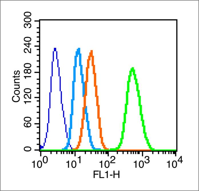
. Overlay histogram showing RAW264.7 cells stained with A00153-4 (Blue line). To facilitate intracellular staining, cells were fixed with 4% paraformaldehyde and permeabilized with permeabilization buffer. The cells were blocked with 10% normal goat serum. And then incubated with rabbit anti-Nanog Antibody (A00153-4, 1 microg/1x106 cells) for 30 min at 20°C. DyLight®488 conjugated goat anti-rabbit IgG (BA1127, 5-10 microg/1x106 cells) was used as secondary antibody for 30 minutes at 20°C. Isotype control antibody (Green line) was rabbit IgG (1 microg/1x106) used under the same conditions. Unlabelled sample without incubation with primary antibody and secondary antibody (Red line) was used as a blank control.")
Figure 1. Western blot analysis of Nanog using anti-Nanog antibody (A00153-4). Electrophoresis was performed on a 5-20% SDS-PAGE gel at 70V (Stacking gel) / 90V (Resolving gel) for 2-3 hours. The sample well of each lane was loaded with 30 ug of sample under reducing conditions. Lane 1: rat testis tissue lysates, Lane 2: rat C6 whole cell lysates, Lane 3: rat PC-12 whole cell lysates, Lane 4: mouse testis tissue lysates, Lane 5: mouse RAW264.7 whole cell lysates. After electrophoresis, proteins were transferred to a nitrocellulose membrane at 150 mA for 50-90 minutes. Blocked the membrane with 5% non-fat milk/TBS for 1.5 hour at RT. The membrane was incubated with rabbit anti-Nanog antigen affinity purified polyclonal antibody (Catalog # A00153-4) at 0.5 microg/mL overnight at 4°C, then washed with TBS-0.1%Tween 3 times with 5 minutes each and probed with a goat anti-rabbit IgG-HRP secondary antibody at a dilution of 1:5000 for 1.5 hour at RT. The signal is developed using an Enhanced Chemiluminescent detection (ECL) kit (Catalog # EK1002) with Tanon 5200 system. A specific band was detected for Nanog at approximately 37 kDa. The expected band size for Nanog is at 37 kDa.
Anti-Nanog Antibody Picoband(r)
A00153-4-CARRIER-FREE
ApplicationsFlow Cytometry, ImmunoFluorescence, Western Blot, ELISA, ImmunoCytoChemistry
Product group Antibodies
ReactivityMouse, Rat
TargetNanog
Overview
- SupplierBoster Bio
- Product NameAnti-Nanog Antibody Picoband(r)
- Delivery Days Customer9
- ApplicationsFlow Cytometry, ImmunoFluorescence, Western Blot, ELISA, ImmunoCytoChemistry
- CertificationResearch Use Only
- ClonalityPolyclonal
- Concentration500 ug/ml
- Gene ID71950
- Target nameNanog
- Target descriptionNanog homeobox
- Target synonyms2410002E02Rik, ENK, Stm1, ecat4, homeobox protein NANOG, ES cell-associated protein 4, early embryo specific expression NK family, early embryo specific expression NK-type homeobox protein, homeobox transcription factor Nanog
- HostRabbit
- IsotypeIgG
- Protein IDQ80Z64
- Protein NameHomeobox protein NANOG
- Scientific DescriptionBoster Bio Anti-Nanog Antibody Picoband® catalog # A00153-4. Tested in ELISA, Flow Cytometry, IF, ICC, WB applications. This antibody reacts with Mouse, Rat. The brand Picoband indicates this is a premium antibody that guarantees superior quality, high affinity, and strong signals with minimal background in Western blot applications. Only our best-performing antibodies are designated as Picoband, ensuring unmatched performance.
- ReactivityMouse, Rat
- Storage Instruction-20°C,2°C to 8°C
- UNSPSC12352203
Related products
Product group Antibodies
References
Nanog Polyclonal AntibodyBS-0829R
ApplicationsFlow Cytometry, ImmunoFluorescence, Western Blot, ELISA, ImmunoCytoChemistry, ImmunoHistoChemistry, ImmunoHistoChemistry Frozen, ImmunoHistoChemistry Paraffin
ReactivityHuman, Mouse, Rat
TargetNanog
- SizePrice
Product group Antibodies
References

Goat anti-NANOGEB06860
ApplicationsImmunoFluorescence, Western Blot, ELISA, ImmunoHistoChemistry
ReactivityCanine, Human, Porcine
TargetNanog
- SizePrice