


Immunohistochemical staining of human small intestine shows strong cytoplasmic positivity in Paneth cells.
Immunohistochemical staining of human small intestine shows strong cytoplasmic positivity in Paneth cells.
Anti-NCF1 Antibody
HPA052095
ApplicationsImmunoHistoChemistry
Product group Antibodies
ReactivityHuman
TargetNCF1
Overview
- SupplierAtlas Antibodies
- Product NameAnti-NCF1 Antibody
- Delivery Days Customer4
- ApplicationsImmunoHistoChemistry
- CertificationResearch Use Only
- ClonalityPolyclonal
- ConjugateUnconjugated
- Gene ID653361
- Target nameNCF1
- Target descriptionneutrophil cytosolic factor 1
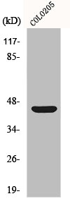
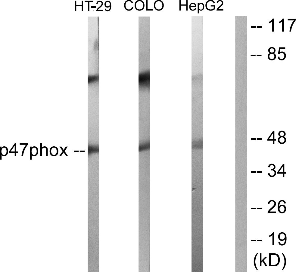
- Target synonymsCGD1, NCF-1, NCF-47K, NCF1A, NOXO2, SH3PXD1A, p47-phox, p47phox, neutrophil cytosol factor 1, 47 kDa autosomal chronic granulomatous disease protein, 47 kDa neutrophil oxidase factor, NADPH oxidase organizer 2, SH3 and PX domain-containing protein 1A, neutrophil NADPH oxidase factor 1, neutrophil cytosolic factor 1, (chronic granulomatous disease, autosomal 1), nox organizer 2, nox-organizing protein 2
- HostRabbit
- IsotypeIgG
- Protein IDP14598
- Protein NameNeutrophil cytosol factor 1
- Scientific DescriptionRecombinant Protein Epitope Signature Tag (PrEST) antigen sequence
- ReactivityHuman
- Storage Instruction-20°C,2°C to 8°C
- UNSPSC41116161
Datasheet
MSDS
Related products
Product group Antibodies
ReactivityHuman
TargetNCF1
- SizePrice
Product group Antibodies
NCF1 AntibodyCSB-PA003664
ApplicationsImmunoFluorescence, Western Blot, ELISA, ImmunoHistoChemistry
ReactivityHuman, Monkey
TargetNCF1
- SizePrice
Product group Antibodies
Anti-NCF1 Antibody Picoband(r)A01586-1-CARRIER-FREE
ApplicationsWestern Blot, ELISA
ReactivityHuman, Mouse, Rat
TargetNCF1
- SizePrice
Product group Antibodies
Anti-NCF1 Antibody144-01148
ApplicationsImmunoPrecipitation, Western Blot
ReactivityHuman, Mouse, Rat
TargetNCF1
- SizePrice
Product group Antibodies
Anti-p47 phox AntibodyA95329
ApplicationsWestern Blot, ELISA, ImmunoHistoChemistry
ReactivityHuman, Mouse, Rat
- SizePrice
Product group Antibodies

ApplicationsWestern Blot, ELISA
- SizePrice

Product group Antibodies
Anti-NCF1 AntibodyHPA047836
ApplicationsImmunoHistoChemistry
ReactivityHuman
TargetNCF1
- SizePrice