

Neutrophil Cytosol Factor 1 (Phospho-Ser304) Antibody
ABX012510
ApplicationsWestern Blot, ELISA
Product group Antibodies
Overview
- SupplierAbbexa
- Product NameNeutrophil Cytosol Factor 1 (Phospho-Ser304) Antibody
- Delivery Days Customer12
- ApplicationsWestern Blot, ELISA
- CertificationResearch Use Only
- ClonalityPolyclonal
- ConjugateUnconjugated
- HostRabbit
- UNSPSC12352203
Related products
Product group Antibodies
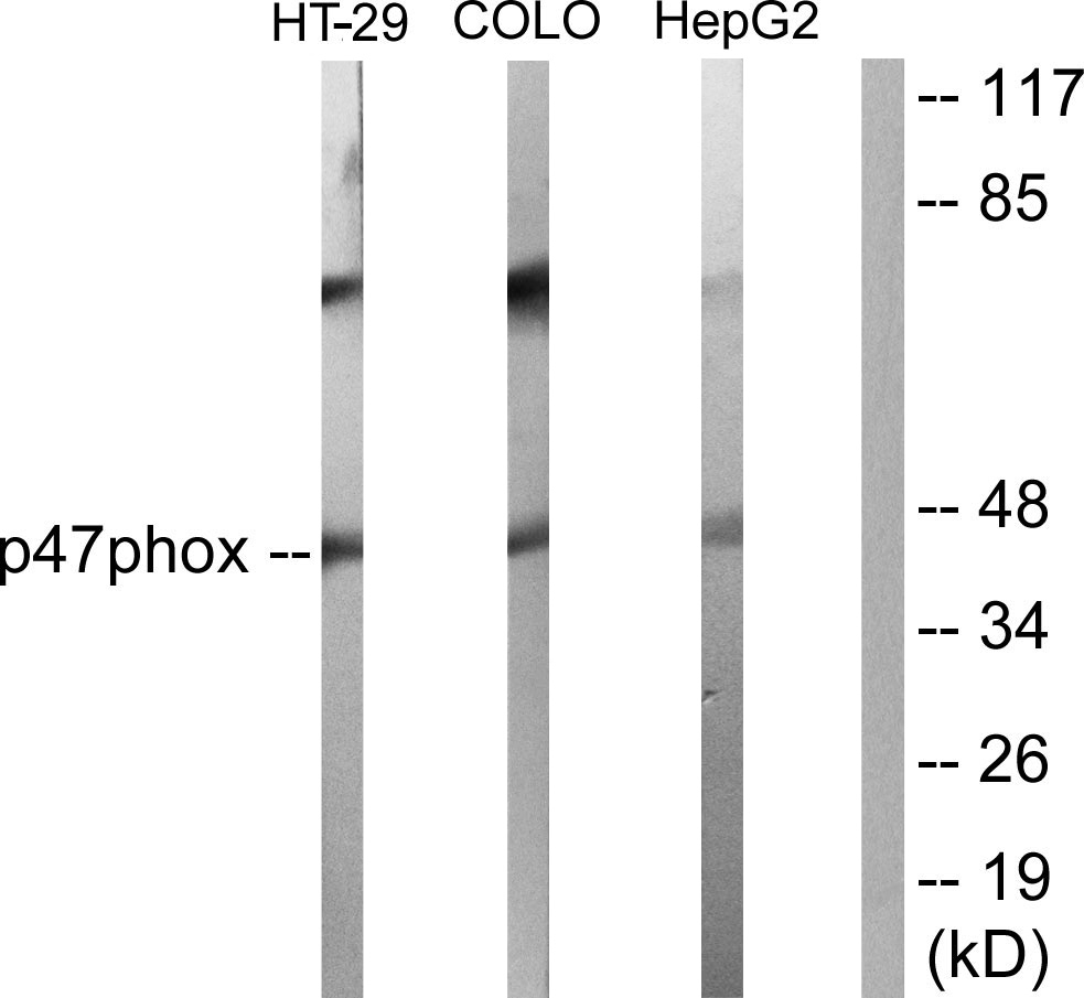
Anti-p47 phox AntibodyA95329
ApplicationsWestern Blot, ELISA, ImmunoHistoChemistry
ReactivityHuman, Mouse, Rat
- SizePrice
Product group Antibodies
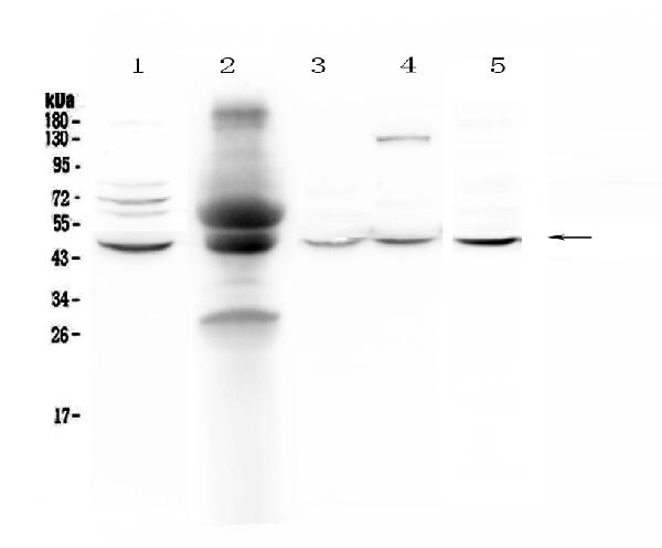
Anti-NCF1 Antibody144-01148
ApplicationsImmunoPrecipitation, Western Blot
ReactivityHuman, Mouse, Rat
TargetNCF1
- SizePrice
Product group Antibodies
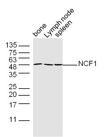
Anti-NCF1 Antibody Picoband(r)A01586-1-CARRIER-FREE
ApplicationsWestern Blot, ELISA
ReactivityHuman, Mouse, Rat
TargetNCF1
- SizePrice
Product group Antibodies
References
NCF1 Polyclonal AntibodyBS-3886R
ApplicationsFlow Cytometry, ImmunoFluorescence, Western Blot, ELISA, ImmunoCytoChemistry, ImmunoHistoChemistry, ImmunoHistoChemistry Paraffin
ReactivityCanine, Human, Mouse, Rat
TargetNCF1
- SizePrice
Product group Antibodies
NCF1 AntibodyCSB-PA003664
ApplicationsImmunoFluorescence, Western Blot, ELISA, ImmunoHistoChemistry
ReactivityHuman, Monkey
TargetNCF1
- SizePrice
Product group Antibodies
NCF1 Polyclonal AntibodyCAC14742
ApplicationsImmunoFluorescence, Western Blot, ELISA, ImmunoHistoChemistry
ReactivityMouse
TargetNCF1
- SizePrice
Product group Antibodies
NCF1 antibodyGTX102957
ApplicationsImmunoFluorescence, Western Blot, ImmunoCytoChemistry, ImmunoHistoChemistry, ImmunoHistoChemistry Paraffin
ReactivityHuman
TargetNCF1
- SizePrice
Product group Antibodies

ApplicationsImmunoFluorescence, Western Blot, ImmunoCytoChemistry, ImmunoHistoChemistry, ImmunoHistoChemistry Paraffin
ReactivityBovine, Canine, Human, Monkey, Mouse, Sheep
TargetNCF1
- SizePrice

Product group Antibodies
Anti-NCF1 AntibodyHPA047836
ApplicationsImmunoHistoChemistry
ReactivityHuman
TargetNCF1
- SizePrice