


Immunohistochemical staining of human skeletal muscle shows moderate cytoplasmic positivity in myocytes.
Immunohistochemical staining of human skeletal muscle shows moderate cytoplasmic positivity in myocytes.
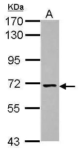
Anti-NCLN Antibody
HPA006625
ApplicationsImmunoHistoChemistry
Product group Antibodies
ReactivityHuman
TargetNCLN
Overview
- SupplierAtlas Antibodies
- Product NameAnti-NCLN Antibody
- Delivery Days Customer4
- ApplicationsImmunoHistoChemistry
- CertificationResearch Use Only
- ClonalityPolyclonal
- ConjugateUnconjugated
- Gene ID56926
- Target nameNCLN
- Target descriptionnicalin
- Target synonymsNET59, BOS complex subunit NCLN, nicalin homolog, nicastrin-like protein
- HostRabbit
- IsotypeIgG
- Protein IDQ969V3
- Protein NameBOS complex subunit NCLN
- Scientific DescriptionRecombinant Protein Epitope Signature Tag (PrEST) antigen sequence
- ReactivityHuman
- Storage Instruction-20°C,2°C to 8°C
- UNSPSC41116161
Datasheet
MSDS
Related products

Product group Antibodies
Anti-NCLN Antibody Picoband(r)A12284-2-CARRIER-FREE
ApplicationsFlow Cytometry, ImmunoFluorescence, Western Blot, ELISA, ImmunoCytoChemistry, ImmunoHistoChemistry
ReactivityHuman, Mouse, Rat
TargetNCLN
- SizePrice

Product group Antibodies
Anti-NCLN AntibodyA98017
ApplicationsWestern Blot, ELISA
ReactivityHuman, Mouse, Rat
- SizePrice
Product group Antibodies

NCLN Antibody (aa375-425)LS-C763133
ApplicationsImmunoPrecipitation, Western Blot
ReactivityHuman, Mouse
TargetNCLN
- SizePrice
Product group Antibodies
NCLN AntibodyCSB-PA040031
ApplicationsWestern Blot, ELISA, ImmunoHistoChemistry
ReactivityHuman, Mouse, Rat
TargetNCLN
- SizePrice
Product group Antibodies
NCLN antibody [N2C2], InternalGTX118355
ApplicationsWestern Blot
ReactivityHuman
TargetNCLN
- SizePrice