


Immunofluorescent staining of human cell line HeLa shows localization to nucleoli, cytosol & the Golgi apparatus.
Immunofluorescent staining of human cell line HeLa shows localization to nucleoli, cytosol & the Golgi apparatus.
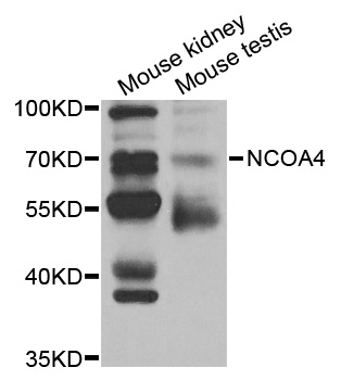
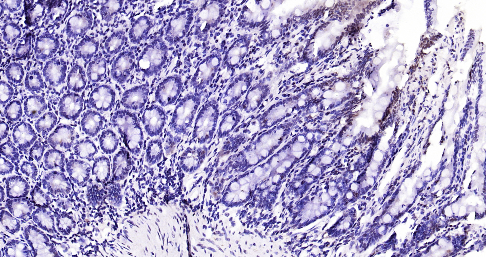
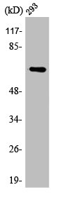
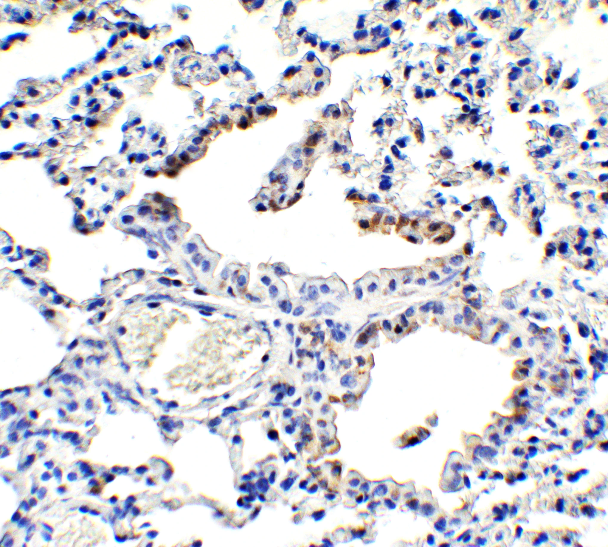
Anti-NCOA4 Antibody
HPA051260
ApplicationsImmunoCytoChemistry
Product group Antibodies
ReactivityHuman
TargetNCOA4
Overview
- SupplierAtlas Antibodies
- Product NameAnti-NCOA4 Antibody
- Delivery Days Customer4
- ApplicationsImmunoCytoChemistry
- CertificationResearch Use Only
- ClonalityPolyclonal
- ConjugateUnconjugated
- Gene ID8031
- Target nameNCOA4
- Target descriptionnuclear receptor coactivator 4
- Target synonymsARA70, ELE1, PTC3, RFG, nuclear receptor coactivator 4, 70 kDa AR-activator, 70 kDa androgen receptor coactivator, NCoA-4, RET-activating gene ELE1, androgen receptor-associated protein of 70 kDa, ferritin cargo receptor NCOA4, ret fused
- HostRabbit
- IsotypeIgG
- Protein IDQ13772
- Protein NameNuclear receptor coactivator 4
- Scientific DescriptionRecombinant Protein Epitope Signature Tag (PrEST) antigen sequence
- ReactivityHuman
- Storage Instruction-20°C,2°C to 8°C
- UNSPSC41116161
Datasheet
MSDS
Related products
Product group Antibodies
Anti-NCOA4 AntibodyA37644
ApplicationsWestern Blot, ImmunoHistoChemistry
ReactivityHuman
- SizePrice

Product group Antibodies
Anti-NCOA4 Antibody Picoband(r)A04368-3-CARRIER-FREE
ApplicationsFlow Cytometry, Western Blot, ELISA
ReactivityHuman, Mouse, Rat
TargetNCOA4
- SizePrice
Product group Antibodies
Anti-NCOA4 Antibody144-61423
ApplicationsWestern Blot, ImmunoHistoChemistry
ReactivityHuman, Mouse, Rat
TargetNCOA4
- SizePrice
Product group Antibodies

ARA70 / NCOA4 AntibodyLS-C830531
ApplicationsELISA, ImmunoHistoChemistry
ReactivityHuman
TargetNCOA4
- SizePrice
Product group Antibodies
NCOA4 Polyclonal AntibodyBS-19051R
ApplicationsImmunoFluorescence, ELISA, ImmunoHistoChemistry, ImmunoHistoChemistry Frozen, ImmunoHistoChemistry Paraffin
ReactivityBovine, Equine, Human, Mouse, Porcine, Rat, Sheep
TargetNCOA4
- SizePrice
Product group Antibodies
NCOA4 AntibodyCSB-PA000917
ApplicationsWestern Blot, ELISA, ImmunoHistoChemistry
ReactivityHuman, Mouse, Rat
TargetNCOA4
- SizePrice
Product group Antibodies
NCOA4 antibodyGTX04250
ApplicationsImmunoFluorescence, Western Blot, ELISA, ImmunoCytoChemistry, ImmunoHistoChemistry, ImmunoHistoChemistry Paraffin
ReactivityHuman, Mouse, Rat
TargetNCOA4
- SizePrice

Product group Antibodies
Anti-NCOA4 AntibodyHPA065208
ApplicationsImmunoHistoChemistry
ReactivityHuman
TargetNCOA4
- SizePrice