


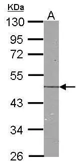
Figure 1. Western blot analysis of NDRG1 using anti-NDRG1 antibody (A01327-1). Electrophoresis was performed on a 5-20% SDS-PAGE gel at 70V (Stacking gel) / 90V (Resolving gel) for 2-3 hours. The sample well of each lane was loaded with 50ug of sample under reducing conditions. Lane 1: human HEK293 whole cell lysates, Lane 2: human U-87MG whole cell lysates. After Electrophoresis, proteins were transferred to a Nitrocellulose membrane at 150mA for 50-90 minutes. Blocked the membrane with 5% Non-fat Milk/ TBS for 1.5 hour at RT. The membrane was incubated with rabbit anti-NDRG1 antigen affinity purified polyclonal antibody (Catalog # A01327-1) at 0.5 microg/mL overnight at 4°C, then washed with TBS-0.1%Tween 3 times with 5 minutes each and probed with a goat anti-rabbit IgG-HRP secondary antibody at a dilution of 1:5000 for 1.5 hour at RT. The signal is developed using an Enhanced Chemiluminescent detection (ECL) kit (Catalog # EK1002) with Tanon 5200 system. A specific band was detected for NDRG1 at approximately 46KD. The expected band size for NDRG1 is at 46KD.
. Overlay histogram showing PC-3 cells stained with A01327-1 (Blue line). To facilitate intracellular staining, cells were fixed with 4% paraformaldehyde and permeabilized with permeabilization buffer. The cells were blocked with 10% normal goat serum. And then incubated with rabbit anti-NDRG1 Antibody (A01327-1, 1microg/1x106 cells) for 30 min at 20°C. DyLight®488 conjugated goat anti-rabbit IgG (BA1127, 5-10microg/1x106 cells) was used as secondary antibody for 30 minutes at 20°C. Isotype control antibody (Green line) was rabbit IgG (1microg/1x106) used under the same conditions. Unlabelled sample without incubation with primary antibody and secondary antibody (Red line) was used as a blank control.")
Figure 1. Western blot analysis of NDRG1 using anti-NDRG1 antibody (A01327-1). Electrophoresis was performed on a 5-20% SDS-PAGE gel at 70V (Stacking gel) / 90V (Resolving gel) for 2-3 hours. The sample well of each lane was loaded with 50ug of sample under reducing conditions. Lane 1: human HEK293 whole cell lysates, Lane 2: human U-87MG whole cell lysates. After Electrophoresis, proteins were transferred to a Nitrocellulose membrane at 150mA for 50-90 minutes. Blocked the membrane with 5% Non-fat Milk/ TBS for 1.5 hour at RT. The membrane was incubated with rabbit anti-NDRG1 antigen affinity purified polyclonal antibody (Catalog # A01327-1) at 0.5 microg/mL overnight at 4°C, then washed with TBS-0.1%Tween 3 times with 5 minutes each and probed with a goat anti-rabbit IgG-HRP secondary antibody at a dilution of 1:5000 for 1.5 hour at RT. The signal is developed using an Enhanced Chemiluminescent detection (ECL) kit (Catalog # EK1002) with Tanon 5200 system. A specific band was detected for NDRG1 at approximately 46KD. The expected band size for NDRG1 is at 46KD.
Anti-NDRG1 Antibody Picoband(r)
A01327-1-CARRIER-FREE
ApplicationsFlow Cytometry, Western Blot, ELISA
Product group Antibodies
ReactivityHuman
TargetNDRG1
Overview
- SupplierBoster Bio
- Product NameAnti-NDRG1 Antibody Picoband(r)
- Delivery Days Customer9
- ApplicationsFlow Cytometry, Western Blot, ELISA
- CertificationResearch Use Only
- ClonalityPolyclonal
- Concentration500 ug/ml
- Gene ID10397
- Target nameNDRG1
- Target descriptionN-myc downstream regulated 1
- Target synonymsCAP43, CMT4D, DRG-1, DRG1, GC4, HMSNL, NDR1, NMSL, PROXY1, RIT42, RTP, TARG1, TDD5, protein NDRG1, N-myc downstream-regulated gene 1 protein, differentiation-related gene 1 protein, nickel-specific induction protein Cap43, protein regulated by oxygen-1, reducing agents and tunicamycin-responsive protein
- HostRabbit
- IsotypeIgG
- Protein IDQ92597
- Protein NameProtein NDRG1
- Scientific DescriptionBoster Bio Anti-NDRG1 Antibody Picoband® catalog # A01327-1. Tested in ELISA, Flow Cytometry, WB applications. This antibody reacts with Human. The brand Picoband indicates this is a premium antibody that guarantees superior quality, high affinity, and strong signals with minimal background in Western blot applications. Only our best-performing antibodies are designated as Picoband, ensuring unmatched performance.
- ReactivityHuman
- Storage Instruction-20°C,2°C to 8°C
- UNSPSC12352203
Related products
Product group Antibodies
NDRG1 AntibodyCSB-PA189423
ApplicationsWestern Blot, ELISA, ImmunoHistoChemistry
ReactivityHuman, Mouse, Rat
TargetNDRG1
- SizePrice
Product group Antibodies
Anti-NDRG1 AntibodyA30038
ApplicationsImmunoFluorescence, Western Blot, ImmunoHistoChemistry
ReactivityHuman, Mouse, Rat
- SizePrice
Product group Antibodies

Goat anti-NDRG1, BiotinylatedEB05707-B
ApplicationsWestern Blot, ELISA, ImmunoHistoChemistry
ReactivityHuman
TargetNDRG1
- SizePrice

Product group Antibodies
Anti-NDRG1 AntibodyHPA006881
ApplicationsWestern Blot, ImmunoCytoChemistry, ImmunoHistoChemistry
ReactivityHuman, Rat
TargetNDRG1
- SizePrice
Product group Antibodies

NDRG1 AntibodyLS-C405949
ApplicationsWestern Blot, ELISA, ImmunoHistoChemistry
ReactivityHuman, Mouse, Rat
TargetNDRG1
- SizePrice
Product group Antibodies
NDRG1 Recombinant Antibody, Biotin ConjugatedBSM-61629R-BIOTIN
ApplicationsImmunoPrecipitation, Western Blot, ImmunoHistoChemistry, ImmunoHistoChemistry Frozen, ImmunoHistoChemistry Paraffin
ReactivityHuman, Mouse, Rat
TargetNDRG1
- SizePrice
Product group Antibodies
NDRG1 Polyclonal AntibodyCAC15826
ApplicationsImmunoFluorescence, Western Blot, ELISA, ImmunoHistoChemistry
TargetNDRG1
- SizePrice
Product group Antibodies
NDRG1 antibodyGTX100537
ApplicationsWestern Blot, ImmunoHistoChemistry, ImmunoHistoChemistry Paraffin
ReactivityHuman, Mouse, Rat
TargetNDRG1
- SizePrice
Product group Antibodies
Anti-NDRG1 Antibody144-02142
ApplicationsImmunoFluorescence, Western Blot, ImmunoHistoChemistry
ReactivityHuman, Mouse, Rat
TargetNDRG1
- SizePrice