


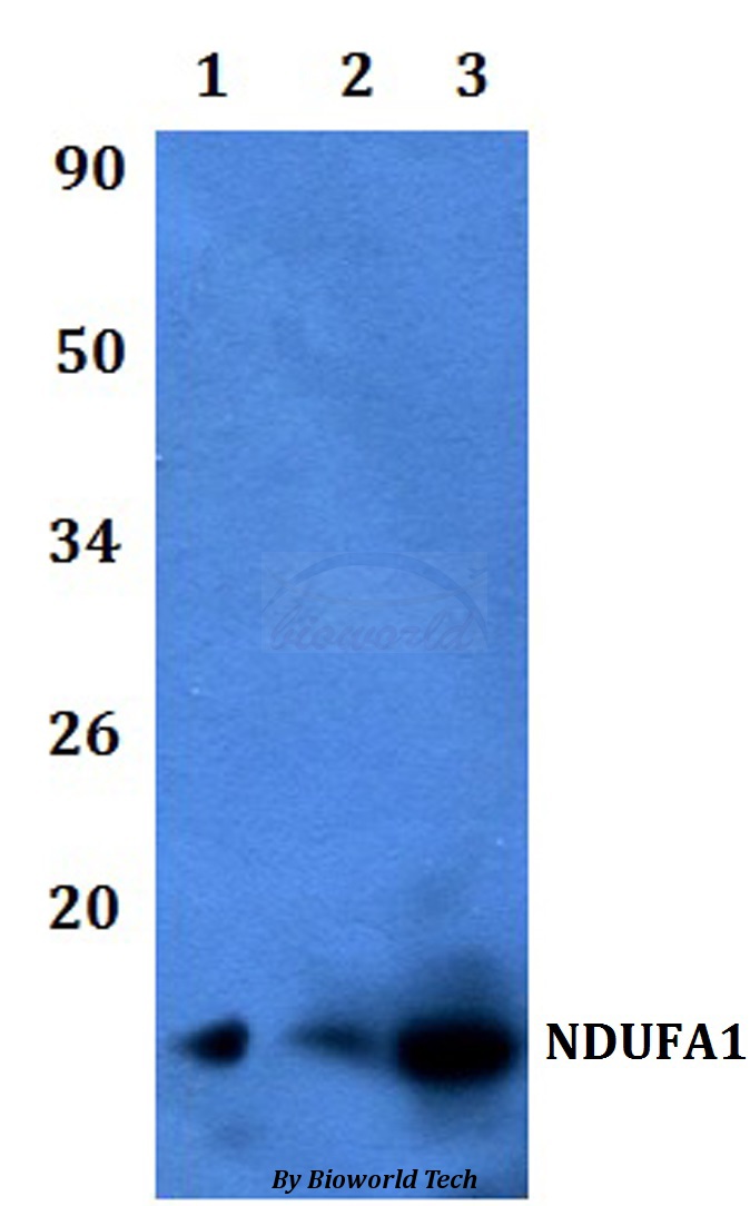
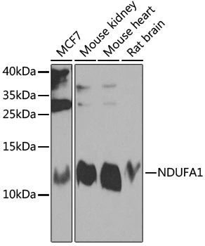
Figure 1. Western blot analysis of NDUFA1 using anti-NDUFA1 antibody (A08224). Electrophoresis was performed on a 5-20% SDS-PAGE gel at 70V (Stacking gel) / 90V (Resolving gel) for 2-3 hours. The sample well of each lane was loaded with 30 ug of sample under reducing conditions. Lane 1: human Hela whole cell lysates, Lane 2: human MCF-7 whole cell lysates, Lane 3: rat kidney tissue lysates, Lane 4: rat liver tissue lysates, Lane 5: rat heart tissue lysates, Lane 6: mouse kidney tissue lysates, Lane 7: mouse liver tissue lysates, Lane 8: mouse heart tissue lysates. After electrophoresis, proteins were transferred to a nitrocellulose membrane at 150 mA for 50-90 minutes. Blocked the membrane with 5% non-fat milk/TBS for 1.5 hour at RT. The membrane was incubated with rabbit anti-NDUFA1 antigen affinity purified polyclonal antibody (Catalog # A08224) at 0.25 microg/mL overnight at 4°C, then washed with TBS-0.1%Tween 3 times with 5 minutes each and probed with a goat anti-rabbit IgG-HRP secondary antibody at a dilution of 1:5000 for 1.5 hour at RT. The signal is developed using an Enhanced Chemiluminescent detection (ECL) kit (Catalog # EK1002) with Tanon 5200 system. A specific band was detected for NDUFA1 at approximately 10 kDa. The expected band size for NDUFA1 is at 8 kDa.
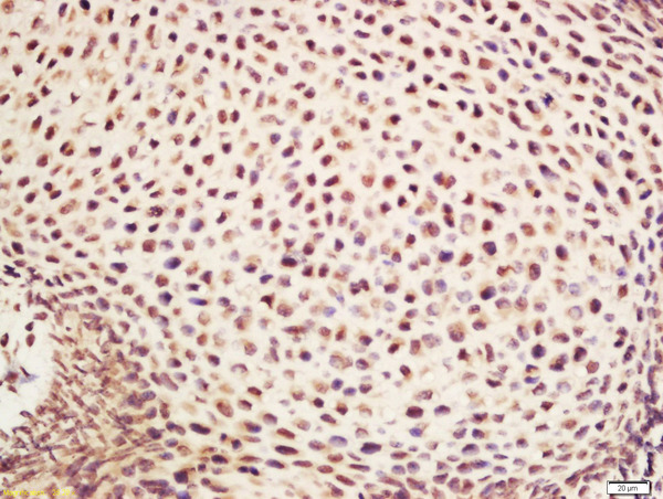
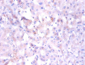
. NDUFA1 was detected in a paraffin-embedded section of human prostate cancer tissue. Heat mediated antigen retrieval was performed in EDTA buffer (pH 8.0, epitope retrieval solution). The tissue section was blocked with 10% goat serum. The tissue section was then incubated with 2 microg/ml rabbit anti-NDUFA1 Antibody (A08224) overnight at 4°C. Peroxidase Conjugated Goat Anti-rabbit IgG was used as secondary antibody and incubated for 30 minutes at 37°C. The tissue section was developed using HRP Conjugated Rabbit IgG Super Vision Assay Kit (Catalog # SV0002) with DAB as the chromogen.")
. NDUFA1 was detected in a paraffin-embedded section of human prostate cancer tissue. Heat mediated antigen retrieval was performed in EDTA buffer (pH 8.0, epitope retrieval solution). The tissue section was blocked with 10% goat serum. The tissue section was then incubated with 2 microg/ml rabbit anti-NDUFA1 Antibody (A08224) overnight at 4°C. Peroxidase Conjugated Goat Anti-rabbit IgG was used as secondary antibody and incubated for 30 minutes at 37°C. The tissue section was developed using HRP Conjugated Rabbit IgG Super Vision Assay Kit (Catalog # SV0002) with DAB as the chromogen.")
Figure 1. Western blot analysis of NDUFA1 using anti-NDUFA1 antibody (A08224). Electrophoresis was performed on a 5-20% SDS-PAGE gel at 70V (Stacking gel) / 90V (Resolving gel) for 2-3 hours. The sample well of each lane was loaded with 30 ug of sample under reducing conditions. Lane 1: human Hela whole cell lysates, Lane 2: human MCF-7 whole cell lysates, Lane 3: rat kidney tissue lysates, Lane 4: rat liver tissue lysates, Lane 5: rat heart tissue lysates, Lane 6: mouse kidney tissue lysates, Lane 7: mouse liver tissue lysates, Lane 8: mouse heart tissue lysates. After electrophoresis, proteins were transferred to a nitrocellulose membrane at 150 mA for 50-90 minutes. Blocked the membrane with 5% non-fat milk/TBS for 1.5 hour at RT. The membrane was incubated with rabbit anti-NDUFA1 antigen affinity purified polyclonal antibody (Catalog # A08224) at 0.25 microg/mL overnight at 4°C, then washed with TBS-0.1%Tween 3 times with 5 minutes each and probed with a goat anti-rabbit IgG-HRP secondary antibody at a dilution of 1:5000 for 1.5 hour at RT. The signal is developed using an Enhanced Chemiluminescent detection (ECL) kit (Catalog # EK1002) with Tanon 5200 system. A specific band was detected for NDUFA1 at approximately 10 kDa. The expected band size for NDUFA1 is at 8 kDa.
Anti-NDUFA1 Antibody Picoband(r)
A08224-DYLIGHT594
ApplicationsWestern Blot, ELISA, ImmunoHistoChemistry
Product group Antibodies
ReactivityHuman, Mouse, Rat
TargetNDUFA1
Overview
- SupplierBoster Bio
- Product NameAnti-NDUFA1 Antibody Picoband(r)
- Delivery Days Customer9
- ApplicationsWestern Blot, ELISA, ImmunoHistoChemistry
- CertificationResearch Use Only
- ClonalityPolyclonal
- Concentration500 ug/ml
- ConjugateOther Conjugate
- Gene ID4694
- Target nameNDUFA1
- Target descriptionNADH:ubiquinone oxidoreductase subunit A1
- Target synonymsCI-MWFE, MC1DN12, MWFE, ZNF183, NADH dehydrogenase [ubiquinone] 1 alpha subcomplex subunit 1, NADH dehydrogenase (ubiquinone) 1 alpha subcomplex, 1, 7.5kDa, NADH-ubiquinone oxidoreductase MWFE subunit, NADH:ubiquinone oxidoreductase (complex 1), complex I MWFE subunit, type I dehydrogenase
- HostRabbit
- Protein IDO15239
- Protein NameNADH dehydrogenase [ubiquinone] 1 alpha subcomplex subunit 1
- Scientific DescriptionBoster Bio Anti-NDUFA1 Antibody Picoband® catalog # A08224. Tested in WB, IHC, ELISA applications. This antibody reacts with Human, Mouse, Rat. The brand Picoband indicates this is a premium antibody that guarantees superior quality, high affinity, and strong signals with minimal background in Western blot applications. Only our best-performing antibodies are designated as Picoband, ensuring unmatched performance.
- ReactivityHuman, Mouse, Rat
- Storage Instruction-20°C,2°C to 8°C
- UNSPSC12352203
Related products
Product group Antibodies
NDUFA1 Polyclonal AntibodyBS-3956R
ApplicationsImmunoFluorescence, ELISA, ImmunoCytoChemistry, ImmunoHistoChemistry, ImmunoHistoChemistry Frozen, ImmunoHistoChemistry Paraffin
ReactivityCanine, Equine, Human, Mouse, Rat
TargetNDUFA1
- SizePrice
Product group Antibodies
Anti-NDUFA1 AntibodyA28656
ApplicationsWestern Blot
ReactivityHuman, Mouse, Rat
- SizePrice
Product group Antibodies
Anti-NDUFA1 Antibody144-08326
ApplicationsWestern Blot
ReactivityHuman, Mouse, Rat
TargetNDUFA1
- SizePrice
Product group Antibodies
NDUFA1 antibodyGTX32740
ApplicationsWestern Blot
ReactivityHuman, Mouse, Rat
TargetNDUFA1
- SizePrice
Product group Antibodies

NDUFA1 AntibodyLS-C401871
ApplicationsELISA, ImmunoHistoChemistry
ReactivityHuman
TargetNDUFA1
- SizePrice

Product group Antibodies
Anti-NDUFA1 AntibodyHPA029768
ApplicationsImmunoCytoChemistry
ReactivityHuman
TargetNDUFA1
- SizePrice
Product group Antibodies
NDUFA1 AntibodyCSB-PA01665A0RB
ApplicationsImmunoFluorescence, ELISA, ImmunoHistoChemistry
ReactivityHuman
TargetNDUFA1
- SizePrice