


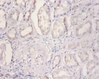
Immunohistochemical staining of human cerebral cortex shows strong cytoplasmic positivity in neuronal cells.
![Lane 1: Marker [kDa] 230, 130, 95, 72, 56, 36, 28, 17, 11. Lane 2: Human cell line RT-4. Lane 3: Human cell line U-251MG sp](https://atlasantibodies.s3.amazonaws.com/images/wb/hpa035933-wb-1.jpg "Lane 1: Marker [kDa] 230, 130, 95, 72, 56, 36, 28, 17, 11. Lane 2: Human cell line RT-4. Lane 3: Human cell line U-251MG sp")
Immunohistochemical staining of human cerebral cortex shows strong cytoplasmic positivity in neuronal cells.
Anti-NDUFA2-25ul
HPA035933
ApplicationsWestern Blot, ImmunoHistoChemistry
Product group Antibodies
ReactivityHuman
Overview
- SupplierAtlas Antibodies
- Product NameAnti-NDUFA2-25ul
- Delivery Days Customer6
- ApplicationsWestern Blot, ImmunoHistoChemistry
- Applications SupplierWB, IHC
- CertificationResearch Use Only
- ClonalityPolyclonal
- Concentration0.1
- ConjugateUnconjugated
- HostRabbit
- IsotypeIgG
- Protein IDO43678
- Protein NameNADH dehydrogenase [ubiquinone] 1 alpha subcomplex subunit 2
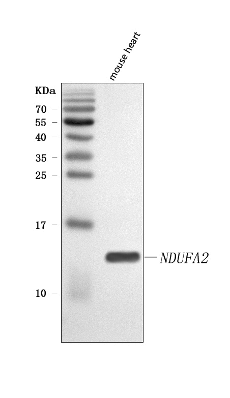
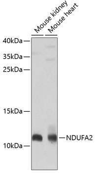
- Scientific DescriptionRabbit Polyclonal Anti-NDUFA2 Antibody against Human NADH dehydrogenase (ubiquinone) 1 alpha subcomplex, 2, 8kDa. Validated for Immunohistochemistry and Western Blot
- ReactivityHuman
- Storage InstructionStore at +4°C for short term storage. Long time storage is recommended at -20°C.
- UNSPSC12352203
Datasheet
MSDS
Related products
Product group Antibodies
Anti-NDUFA2 Antibody Picoband(r)A10396-1-CARRIER-FREE
ApplicationsWestern Blot, ELISA
ReactivityHuman, Mouse
TargetNDUFA2
- SizePrice
Product group Antibodies
Anti-NDUFA2 AntibodyA12213
ApplicationsImmunoFluorescence, Western Blot, ImmunoCytoChemistry
ReactivityHuman, Mouse, Rat
- SizePrice
Product group Antibodies
Anti-NDUFA2 Antibody144-08136
ApplicationsWestern Blot, ImmunoHistoChemistry
ReactivityHuman, Mouse
TargetNDUFA2
- SizePrice
Product group Antibodies

NDUFA2 AntibodyLS-C830294
ApplicationsELISA, ImmunoHistoChemistry
ReactivityHuman
TargetNDUFA2
- SizePrice
Product group Antibodies
NDUFA2 AntibodyCSB-PA02989A0RB
ApplicationsELISA, ImmunoHistoChemistry
ReactivityHuman
TargetNDUFA2
- SizePrice
Product group Antibodies
NDUFA2 antibodyGTX32741
ApplicationsImmunoFluorescence, Western Blot, ImmunoCytoChemistry, ImmunoHistoChemistry, ImmunoHistoChemistry Paraffin
ReactivityHuman, Mouse
TargetNDUFA2
- SizePrice
Product group Antibodies
Anti-NDUFA2 AntibodyCAB8136
ApplicationsImmunoFluorescence, Western Blot, ELISA, ImmunoCytoChemistry
ReactivityHuman
TargetNDUFA2
- SizePrice