

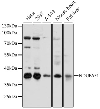
Anti-NDUFAF1 Antibody
A89629
ApplicationsWestern Blot
Product group Antibodies
ReactivityHuman, Mouse, Rat
Overview
- SupplierAntibodies.com
- Product NameAnti-NDUFAF1 Antibody
- Delivery Days Customer7
- ApplicationsWestern Blot
- CertificationResearch Use Only
- ClonalityPolyclonal
- ConjugateUnconjugated
- HostRabbit
- IsotypeIgG
- Scientific DescriptionRabbit polyclonal antibody to NDUFAF1.
- ReactivityHuman, Mouse, Rat
- UNSPSC12352203
Related products
Product group Antibodies
Anti-NDUFAF1 (C-term) Antibody102-20604
ApplicationsWestern Blot, ImmunoHistoChemistry, ImmunoHistoChemistry Paraffin
TargetNDUFAF1
- SizePrice

Product group Antibodies
Anti-NDUFAF1 Antibody Picoband(r)A10145-CARRIER-FREE
ApplicationsImmunoFluorescence, Western Blot, ELISA, ImmunoCytoChemistry, ImmunoHistoChemistry
ReactivityHuman, Mouse, Rat
TargetNDUFAF1
- SizePrice
Product group Antibodies
NDUFAF1 Recombinant Antibody, AbBy Fluor-647 ConjugatedBSM-62045R-BF647
ApplicationsImmunoFluorescence, Western Blot
ReactivityHuman
TargetNDUFAF1
- SizePrice
Product group Antibodies
NDUFAF1 AntibodyCSB-PA226934
ApplicationsELISA, ImmunoHistoChemistry
ReactivityHuman, Mouse
TargetNDUFAF1
- SizePrice
Product group Antibodies

NDUFAF1 / CIA30 AntibodyLS-C401872
ApplicationsWestern Blot, ELISA
ReactivityHuman, Mouse
TargetNDUFAF1
- SizePrice

Product group Antibodies
Anti-NDUFAF1 AntibodyHPA039933
ApplicationsImmunoCytoChemistry, ImmunoHistoChemistry
ReactivityHuman
TargetNDUFAF1
- SizePrice