


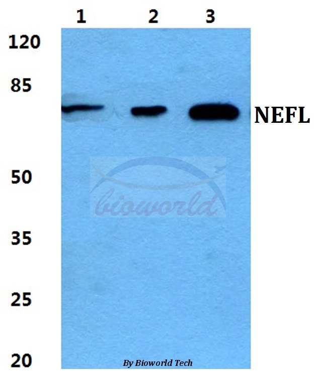
Figure 1. Western blot analysis of NEFL/NF-L using anti-NEFL/NF-L antibody (A02482-1). Electrophoresis was performed on a 5-20% SDS-PAGE gel at 70V (Stacking gel) / 90V (Resolving gel) for 2-3 hours. The sample well of each lane was loaded with 50ug of sample under reducing conditions. Lane 1: human SH-SY5Y whole cell lysates, Lane 2: rat brain tissue lysates, Lane 3: mouse brain tissue lysates. After Electrophoresis, proteins were transferred to a Nitrocellulose membrane at 150mA for 50-90 minutes. Blocked the membrane with 5% Non-fat Milk/ TBS for 1.5 hour at RT. The membrane was incubated with rabbit anti-NEFL/NF-L antigen affinity purified polyclonal antibody (Catalog # A02482-1) at 0.5 microg/mL overnight at 4°C, then washed with TBS-0.1%Tween 3 times with 5 minutes each and probed with a goat anti-rabbit IgG-HRP secondary antibody at a dilution of 1:5000 for 1.5 hour at RT. The signal is developed using an Enhanced Chemiluminescent detection (ECL) kit (Catalog # EK1002) with Tanon 5200 system. A specific band was detected for NEFL/NF-L at approximately 72KD. The expected band size for NEFL/NF-L is at 72KD.
. NEFL/NF-L was detected in paraffin-embedded section of mouse spinal cord tissue. Heat mediated antigen retrieval was performed in EDTA buffer (pH8.0, epitope retrieval solution). The tissue section was blocked with 10% goat serum. The tissue section was then incubated with 2microg/ml rabbit anti-NEFL/NF-L Antibody (A02482-1) overnight at 4°C. Biotinylated goat anti-rabbit IgG was used as secondary antibody and incubated for 30 minutes at 37°C. The tissue section was developed using Strepavidin-Biotin-Complex (SABC) (Catalog # SA1022) with DAB as the chromogen.")
. Overlay histogram showing 293T cells stained with A02482-1 (Blue line). To facilitate intracellular staining, cells were fixed with 4% paraformaldehyde and permeabilized with permeabilization buffer. The cells were blocked with 10% normal goat serum. And then incubated with rabbit anti-NEFL/NF-L Antibody (A02482-1, 1microg/1x106 cells) for 30 min at 20°C. DyLight®488 conjugated goat anti-rabbit IgG (BA1127, 5-10microg/1x106 cells) was used as secondary antibody for 30 minutes at 20°C. Isotype control antibody (Green line) was rabbit IgG (1microg/1x106) used under the same conditions. Unlabelled sample without incubation with primary antibody and secondary antibody (Red line) was used as a blank control.")
. NEFL/NF-L was detected in paraffin-embedded section of rat spinal cord tissue. Heat mediated antigen retrieval was performed in EDTA buffer (pH8.0, epitope retrieval solution). The tissue section was blocked with 10% goat serum. The tissue section was then incubated with 2microg/ml rabbit anti-NEFL/NF-L Antibody (A02482-1) overnight at 4°C. Biotinylated goat anti-rabbit IgG was used as secondary antibody and incubated for 30 minutes at 37°C. The tissue section was developed using Strepavidin-Biotin-Complex (SABC) (Catalog # SA1022) with DAB as the chromogen.")
. NEFL/NF-L was detected in paraffin-embedded section of mouse spinal cord tissue. Heat mediated antigen retrieval was performed in EDTA buffer (pH8.0, epitope retrieval solution). The tissue section was blocked with 10% goat serum. The tissue section was then incubated with 2microg/ml rabbit anti-NEFL/NF-L Antibody (A02482-1) overnight at 4°C. Biotinylated goat anti-rabbit IgG was used as secondary antibody and incubated for 30 minutes at 37°C. The tissue section was developed using Strepavidin-Biotin-Complex (SABC) (Catalog # SA1022) with DAB as the chromogen.")
. NEFL/NF-L was detected in a paraffin-embedded section of mouse brain tissue. Heat mediated antigen retrieval was performed in EDTA buffer (pH 8.0, epitope retrieval solution). The tissue section was blocked with 10% goat serum. The tissue section was then incubated with 5 microg/mL rabbit anti-NEFL/NF-L Antibody (A02482-1) overnight at 4°C. Biotin conjugated goat anti-rabbit IgG (BA1003) was used as secondary antibody and incubated for 30 minutes at 37°C. The tissue section was developed using Cy3 Conjugated Avidin (BA1037). The section was counterstained with DAPI. Visualize using a fluorescence microscope and filter sets appropriate for the label used.")
. NEFL/NF-L was detected in a paraffin-embedded section of rat brain tissue. Heat mediated antigen retrieval was performed in EDTA buffer (pH 8.0, epitope retrieval solution). The tissue section was blocked with 10% goat serum. The tissue section was then incubated with 5 microg/mL rabbit anti-NEFL/NF-L Antibody (A02482-1) overnight at 4°C. Biotin conjugated goat anti-rabbit IgG (BA1003) was used as secondary antibody and incubated for 30 minutes at 37°C. The tissue section was developed using Cy3 Conjugated Avidin (BA1037). The section was counterstained with DAPI. Visualize using a fluorescence microscope and filter sets appropriate for the label used.")
Figure 1. Western blot analysis of NEFL/NF-L using anti-NEFL/NF-L antibody (A02482-1). Electrophoresis was performed on a 5-20% SDS-PAGE gel at 70V (Stacking gel) / 90V (Resolving gel) for 2-3 hours. The sample well of each lane was loaded with 50ug of sample under reducing conditions. Lane 1: human SH-SY5Y whole cell lysates, Lane 2: rat brain tissue lysates, Lane 3: mouse brain tissue lysates. After Electrophoresis, proteins were transferred to a Nitrocellulose membrane at 150mA for 50-90 minutes. Blocked the membrane with 5% Non-fat Milk/ TBS for 1.5 hour at RT. The membrane was incubated with rabbit anti-NEFL/NF-L antigen affinity purified polyclonal antibody (Catalog # A02482-1) at 0.5 microg/mL overnight at 4°C, then washed with TBS-0.1%Tween 3 times with 5 minutes each and probed with a goat anti-rabbit IgG-HRP secondary antibody at a dilution of 1:5000 for 1.5 hour at RT. The signal is developed using an Enhanced Chemiluminescent detection (ECL) kit (Catalog # EK1002) with Tanon 5200 system. A specific band was detected for NEFL/NF-L at approximately 72KD. The expected band size for NEFL/NF-L is at 72KD.
Anti-NEFL/NF-L Antibody Picoband(r)
A02482-1-CARRIER-FREE
ApplicationsFlow Cytometry, ImmunoFluorescence, Western Blot, ELISA, ImmunoHistoChemistry
Product group Antibodies
ReactivityHuman, Mouse, Rat
TargetNEFL
Overview
- SupplierBoster Bio
- Product NameAnti-NEFL/NF-L Antibody Picoband(r)
- Delivery Days Customer9
- ApplicationsFlow Cytometry, ImmunoFluorescence, Western Blot, ELISA, ImmunoHistoChemistry
- CertificationResearch Use Only
- ClonalityPolyclonal
- Concentration500 ug/ml
- Gene ID4747
- Target nameNEFL
- Target descriptionneurofilament light chain
- Target synonymsCMT1F, CMT2E, CMTDIG, NF-L, NF68, NFL, PPP1R110, neurofilament light polypeptide, light molecular weight neurofilament protein, neurofilament protein, light chain, neurofilament subunit NF-L, neurofilament triplet L protein, neurofilament, light polypeptide 68kDa, protein phosphatase 1, regulatory subunit 110
- HostRabbit
- IsotypeIgG
- Protein IDP07196
- Protein NameNeurofilament light polypeptide
- Scientific DescriptionBoster Bio Anti-NEFL/NF-L Antibody Picoband® catalog # A02482-1. Tested in ELISA, Flow Cytometry, IF, IHC, WB applications. This antibody reacts with Human, Mouse, Rat. The brand Picoband indicates this is a premium antibody that guarantees superior quality, high affinity, and strong signals with minimal background in Western blot applications. Only our best-performing antibodies are designated as Picoband, ensuring unmatched performance.
- ReactivityHuman, Mouse, Rat
- Storage Instruction-20°C,2°C to 8°C
- UNSPSC12352203
Related products
Product group Antibodies
Anti-NEFL AntibodyA28063
ApplicationsWestern Blot
ReactivityHuman, Mouse, Rat
- SizePrice
Product group Antibodies
Anti-NEFL Antibody144-00257
ApplicationsImmunoFluorescence, Western Blot, ImmunoHistoChemistry
ReactivityHuman, Mouse, Rat
TargetNEFL
- SizePrice

Product group Antibodies
Anti-NEFL AntibodyAMAB91314
ApplicationsWestern Blot, ImmunoCytoChemistry, ImmunoHistoChemistry
ReactivityHuman, Mouse, Rat
TargetNEFL
- SizePrice
Product group Antibodies

Anti-Neurofilament light [MS-C2]AB00946-10.0-BT
ApplicationsELISA
ReactivityHuman
TargetNEFL
- SizePrice
Product group Antibodies
NF-L Polyclonal AntibodyBS-0707R
ApplicationsFlow Cytometry, ImmunoFluorescence, Western Blot, ImmunoHistoChemistry, ImmunoHistoChemistry Frozen, ImmunoHistoChemistry Paraffin
ReactivityHuman, Mouse, Rat
TargetNEFL
- SizePrice
Product group Antibodies

Goat anti-NEFL (aa330-343)EB13051
ApplicationsWestern Blot, ELISA
ReactivityBovine, Canine, Human, Mouse, Porcine, Rat
TargetNEFL
- SizePrice
Product group Antibodies
Nefl Polyclonal AntibodyCAC08666
ApplicationsImmunoFluorescence, ImmunoPrecipitation, ELISA, ImmunoHistoChemistry
TargetNEFL
- SizePrice
Product group Antibodies
NEFL AntibodyCSB-PA004743
ApplicationsWestern Blot, ELISA
ReactivityHuman, Mouse, Rat
TargetNEFL
- SizePrice
Product group Antibodies

Mouse anti Neurofilament 70 kDMUB1303P
ApplicationsWestern Blot, ImmunoHistoChemistry, ImmunoHistoChemistry Frozen, ImmunoHistoChemistry Paraffin
ReactivityHuman
TargetNEFL
- SizePrice