


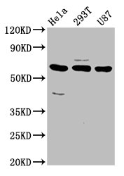
Figure 1. Western blot analysis of NETO1 using anti-NETO1 antibody (A08303-1). Electrophoresis was performed on a 5-20% SDS-PAGE gel at 70V (Stacking gel) / 90V (Resolving gel) for 2-3 hours. The sample well of each lane was loaded with 30 ug of sample under reducing conditions. Lane 1: human RT4 whole cell lysates, Lane 2: human U20S whole cell lysates, Lane 3: human PC-3 whole cell lysates, Lane 4: human SIHA whole cell lysates, Lane 5: rat C6 whole cell lysates, Lane 6: rat NRK whole cell lysates, Lane 7: mouse RM-1 whole cell lysates, Lane 8: mouse SP2/0 whole cell lysates. After electrophoresis, proteins were transferred to a nitrocellulose membrane at 150 mA for 50-90 minutes. Blocked the membrane with 5% non-fat milk/TBS for 1.5 hour at RT. The membrane was incubated with rabbit anti-NETO1 antigen affinity purified polyclonal antibody (Catalog # A08303-1) at 0.5 microg/mL overnight at 4°C, then washed with TBS-0.1%Tween 3 times with 5 minutes each and probed with a goat anti-rabbit IgG-HRP secondary antibody at a dilution of 1:5000 for 1.5 hour at RT. The signal is developed using an Enhanced Chemiluminescent detection (ECL) kit (Catalog # EK1002) with Tanon 5200 system. A specific band was detected for NETO1 at approximately 70 kDa. The expected band size for NETO1 is at 60 kDa.
Figure 1. Western blot analysis of NETO1 using anti-NETO1 antibody (A08303-1). Electrophoresis was performed on a 5-20% SDS-PAGE gel at 70V (Stacking gel) / 90V (Resolving gel) for 2-3 hours. The sample well of each lane was loaded with 30 ug of sample under reducing conditions. Lane 1: human RT4 whole cell lysates, Lane 2: human U20S whole cell lysates, Lane 3: human PC-3 whole cell lysates, Lane 4: human SIHA whole cell lysates, Lane 5: rat C6 whole cell lysates, Lane 6: rat NRK whole cell lysates, Lane 7: mouse RM-1 whole cell lysates, Lane 8: mouse SP2/0 whole cell lysates. After electrophoresis, proteins were transferred to a nitrocellulose membrane at 150 mA for 50-90 minutes. Blocked the membrane with 5% non-fat milk/TBS for 1.5 hour at RT. The membrane was incubated with rabbit anti-NETO1 antigen affinity purified polyclonal antibody (Catalog # A08303-1) at 0.5 microg/mL overnight at 4°C, then washed with TBS-0.1%Tween 3 times with 5 minutes each and probed with a goat anti-rabbit IgG-HRP secondary antibody at a dilution of 1:5000 for 1.5 hour at RT. The signal is developed using an Enhanced Chemiluminescent detection (ECL) kit (Catalog # EK1002) with Tanon 5200 system. A specific band was detected for NETO1 at approximately 70 kDa. The expected band size for NETO1 is at 60 kDa.
Anti-NETO1 Antibody Picoband(r)
A08303-1-CARRIER-FREE
ApplicationsWestern Blot, ELISA
Product group Antibodies
ReactivityHuman, Mouse, Rat
TargetNETO1
Overview
- SupplierBoster Bio
- Product NameAnti-NETO1 Antibody Picoband(r)
- Delivery Days Customer9
- ApplicationsWestern Blot, ELISA
- CertificationResearch Use Only
- ClonalityPolyclonal
- Concentration500 ug/ml
- Gene ID81832
- Target nameNETO1
- Target descriptionneuropilin and tolloid like 1
- Target synonymsBCTL1, BTCL1, neuropilin and tolloid-like protein 1, brain-specific transmembrane protein containing 2 CUB and 1 LDL-receptor class A domains protein 1, neuropilin (NRP) and tolloid (TLL)-like 1
- HostRabbit
- Protein IDQ8TDF5
- Protein NameNeuropilin and tolloid-like protein 1
- Scientific DescriptionBoster Bio Anti-NETO1 Antibody Picoband® catalog # A08303-1. Tested in WB, ELISA applications. This antibody reacts with Human, Mouse, Rat. The brand Picoband indicates this is a premium antibody that guarantees superior quality, high affinity, and strong signals with minimal background in Western blot applications. Only our best-performing antibodies are designated as Picoband, ensuring unmatched performance.
- ReactivityHuman, Mouse, Rat
- Storage Instruction-20°C,2°C to 8°C
- UNSPSC12352203
Related products

Product group Antibodies
Anti-NETO1 AntibodyA49479
ApplicationsWestern Blot, ELISA
ReactivityHuman, Mouse, Rat
- SizePrice
Product group Antibodies

NETO1 AntibodyLS-C674230
ApplicationsImmunoFluorescence, Western Blot, ELISA
ReactivityHuman
TargetNETO1
- SizePrice
Product group Antibodies
NETO1 AntibodyCSB-PA840571LA01HU
ApplicationsImmunoFluorescence, Western Blot, ELISA
ReactivityHuman
TargetNETO1
- SizePrice
Product group Antibodies
NETO1 Polyclonal AntibodyCAC13432
ApplicationsImmunoFluorescence, Western Blot, ELISA
TargetNETO1
- SizePrice
Product group Antibodies
NETO1 antibodyGTX31454
ApplicationsWestern Blot, ELISA, ImmunoHistoChemistry, ImmunoHistoChemistry Paraffin
ReactivityHuman, Mouse, Rat
TargetNETO1
- SizePrice

Product group Antibodies
Anti-NETO1 AntibodyHPA064630
ApplicationsImmunoCytoChemistry
ReactivityHuman
TargetNETO1
- SizePrice
Product group Antibodies
NETO1 AntibodyPACO58348
ApplicationsImmunoFluorescence, Western Blot, ELISA
ReactivityHuman
TargetNETO1
- SizePrice