


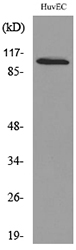
Figure 1. Western blot analysis of NFAT2 using anti-NFAT2 antibody (A00340-1). Electrophoresis was performed on a 5-20% SDS-PAGE gel at 70V (Stacking gel) / 90V (Resolving gel) for 2-3 hours. The sample well of each lane was loaded with 50ug of sample under reducing conditions. Lane 1: mouse thymus tissue lysates, Lane 2: human 22RV1 whole cell lysates. After Electrophoresis, proteins were transferred to a Nitrocellulose membrane at 150mA for 50-90 minutes. Blocked the membrane with 5% Non-fat Milk/ TBS for 1.5 hour at RT. The membrane was incubated with rabbit anti-NFAT2 antigen affinity purified polyclonal antibody (Catalog # A00340-1) at 0.5 ug/mL overnight at 4 then washed with TBS-0.1%Tween 3 times with 5 minutes each and probed with a goat anti-rabbit IgG-HRP secondary antibody at a dilution of 1:10000 for 1.5 hour at RT. The signal is developed using an Enhanced Chemiluminescent detection (ECL) kit (Catalog # EK1002) with Tanon 5200 system. A specific band was detected for NFAT2 at approximately 101KD. The expected band size for NFAT2 is at 101KD.
. Overlay histogram showing Hela cells stained with A00340-1 (Blue line). To facilitate intracellular staining, cells were fixed with 4% paraformaldehyde and permeabilized with permeabilization buffer. The cells were blocked with 10% normal goat serum. And then incubated with rabbit anti-NFATC1 Antibody (A00340-1,1microg/1x106 cells) for 30 min at 20°C. DyLight®488 conjugated goat anti-rabbit IgG (BA1127, 5-10microg/1x106 cells) was used as secondary antibody for 30 minutes at 20°C. Isotype control antibody (Green line) was rabbit IgG (1microg/1x106) used under the same conditions. Unlabelled sample without incubation with primary antibody and secondary antibody (Red line) was used as a blank control.")
. Overlay histogram showing THP-1 cells stained with A00340-1 (Blue line). To facilitate intracellular staining, cells were fixed with 4% paraformaldehyde and permeabilized with permeabilization buffer. The cells were blocked with 10% normal goat serum. And then incubated with rabbit anti-NFATC1 Antibody (A00340-1,1microg/1x106 cells) for 30 min at 20°C. DyLight®488 conjugated goat anti-rabbit IgG (BA1127, 5-10microg/1x106 cells) was used as secondary antibody for 30 minutes at 20°C. Isotype control antibody (Green line) was rabbit IgG (1microg/1x106) used under the same conditions. Unlabelled sample without incubation with primary antibody and secondary antibody (Red line) was used as a blank control.")
. Overlay histogram showing MCF-7 cells stained with A00340-1 (Blue line). To facilitate intracellular staining, cells were fixed with 4% paraformaldehyde and permeabilized with permeabilization buffer. The cells were blocked with 10% normal goat serum. And then incubated with rabbit anti-NFATC1 Antibody (A00340-1,1microg/1x106 cells) for 30 min at 20°C. DyLight®488 conjugated goat anti-rabbit IgG (BA1127, 5-10microg/1x106 cells) was used as secondary antibody for 30 minutes at 20°C. Isotype control antibody (Green line) was rabbit IgG (1microg/1x106) used under the same conditions. Unlabelled sample without incubation with primary antibody and secondary antibody (Red line) was used as a blank control.")
. Overlay histogram showing Jurkat cells stained with A00340-1 (Blue line). To facilitate intracellular staining, cells were fixed with 4% paraformaldehyde and permeabilized with permeabilization buffer. The cells were blocked with 10% normal goat serum. And then incubated with rabbit anti-NFATC1 Antibody (A00340-1,1microg/1x106 cells) for 30 min at 20°C. DyLight®488 conjugated goat anti-rabbit IgG (BA1127, 5-10microg/1x106 cells) was used as secondary antibody for 30 minutes at 20°C. Isotype control antibody (Green line) was rabbit IgG (1microg/1x106) used under the same conditions. Unlabelled sample without incubation with primary antibody and secondary antibody (Red line) was used as a blank control.")
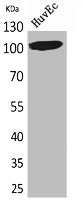
Figure 1. Western blot analysis of NFAT2 using anti-NFAT2 antibody (A00340-1). Electrophoresis was performed on a 5-20% SDS-PAGE gel at 70V (Stacking gel) / 90V (Resolving gel) for 2-3 hours. The sample well of each lane was loaded with 50ug of sample under reducing conditions. Lane 1: mouse thymus tissue lysates, Lane 2: human 22RV1 whole cell lysates. After Electrophoresis, proteins were transferred to a Nitrocellulose membrane at 150mA for 50-90 minutes. Blocked the membrane with 5% Non-fat Milk/ TBS for 1.5 hour at RT. The membrane was incubated with rabbit anti-NFAT2 antigen affinity purified polyclonal antibody (Catalog # A00340-1) at 0.5 ug/mL overnight at 4 then washed with TBS-0.1%Tween 3 times with 5 minutes each and probed with a goat anti-rabbit IgG-HRP secondary antibody at a dilution of 1:10000 for 1.5 hour at RT. The signal is developed using an Enhanced Chemiluminescent detection (ECL) kit (Catalog # EK1002) with Tanon 5200 system. A specific band was detected for NFAT2 at approximately 101KD. The expected band size for NFAT2 is at 101KD.
Anti-NFAT2/NFATC1 Antibody Picoband(r)
A00340-1-CARRIER-FREE
ApplicationsWestern Blot, ELISA, ImmunoHistoChemistry
Product group Antibodies
ReactivityHuman
TargetNFATC1
Overview
- SupplierBoster Bio
- Product NameAnti-NFAT2/NFATC1 Antibody Picoband(r)
- Delivery Days Customer9
- ApplicationsWestern Blot, ELISA, ImmunoHistoChemistry
- CertificationResearch Use Only
- ClonalityPolyclonal
- Concentration500 ug/ml
- Gene ID4772
- Target nameNFATC1
- Target descriptionnuclear factor of activated T cells 1
- Target synonymsNF-ATC, NF-ATc1.2, NFAT2, NFATc, nuclear factor of activated T-cells, cytoplasmic 1, NFAT transcription complex cytosolic component, nuclear factor of activated T-cells 'c', nuclear factor of activated T-cells, cytoplasmic, calcineurin-dependent 1
- HostRabbit
- IsotypeIgG
- Protein IDO95644
- Protein NameNuclear factor of activated T-cells, cytoplasmic 1
- Scientific DescriptionBoster Bio Anti-NFAT2/NFATC1 Antibody Picoband® catalog # A00340-1. Tested in ELISA, IHC, WB applications. This antibody reacts with Human. The brand Picoband indicates this is a premium antibody that guarantees superior quality, high affinity, and strong signals with minimal background in Western blot applications. Only our best-performing antibodies are designated as Picoband, ensuring unmatched performance.
- ReactivityHuman
- Storage Instruction-20°C,2°C to 8°C
- UNSPSC12352203
Related products
Product group Antibodies
Anti-NFATC1 AntibodyA98780
ApplicationsWestern Blot, ELISA
ReactivityHuman, Mouse
- SizePrice
Product group Antibodies
anti-NFATc1, pAb (IG-205)AG-25T-0110
ApplicationsFunctional Assay, ImmunoPrecipitation, Western Blot, ImmunoCytoChemistry, ImmunoHistoChemistry
ReactivityHuman
TargetNFATC1
- SizePrice
Product group Antibodies
Anti-NFATC1 Antibody144-01539
ApplicationsImmunoFluorescence, Western Blot, ImmunoHistoChemistry
ReactivityHuman, Mouse, Rat
TargetNFATC1
- SizePrice
Product group Antibodies
NFAT2 Recombinant Antibody, AbBy Fluor-350 ConjugatedBSM-61483R-BF350
ApplicationsFlow Cytometry, Western Blot
ReactivityHuman
TargetNFATC1
- SizePrice
Product group Antibodies
NFATC1 AntibodyCSB-PA005617
ApplicationsWestern Blot, ELISA
ReactivityHuman, Mouse
TargetNFATC1
- SizePrice
![Various whole cell extracts (30 μg) were separated by 5% SDS-PAGE, and the membrane was blotted with NFAT2 antibody [GT1177] (GTX09510) diluted at 1:1000. The HRP-conjugated anti-rabbit IgG antibody (GTX213110-01) was used to detect the primary antibody.](https://www.genetex.com/upload/website/prouct_img/normal/GTX09510/GTX09510_4000000076_20200410_WB_w_23053123_389.webp)
Product group Antibodies
References
NFAT2 antibody [GT1177]GTX09510
ApplicationsWestern Blot
ReactivityHuman, Mouse
TargetNFATC1
- SizePrice
Product group Antibodies

NFATC1 / NFAT2 AntibodyLS-C331522
ApplicationsImmunoFluorescence, Western Blot, ImmunoHistoChemistry
ReactivityHuman, Mouse
TargetNFATC1
- SizePrice

Product group Antibodies
Anti-NFATC1 AntibodyHPA071732
ApplicationsChIP Chromatin ImmunoPrecipitation, ImmunoCytoChemistry
ReactivityHuman
TargetNFATC1
- SizePrice