


Figure 1. Western blot analysis of NGF using anti-NGF antibody (A00341-1). Electrophoresis was performed on a 5-20% SDS-PAGE gel at 70V (Stacking gel) / 90V (Resolving gel) for 2-3 hours. The sample well of each lane was loaded with 50ug of sample under reducing conditions. Lane 1: human U-87MG whole cell lysates, Lane 2: human 22RV1 whole cell lysates, Lane 3: human A549 whole cell lysates. After Electrophoresis, proteins were transferred to a Nitrocellulose membrane at 150mA for 50-90 minutes. Blocked the membrane with 5% Non-fat Milk/ TBS for 1.5 hour at RT. The membrane was incubated with rabbit anti-NGF antigen affinity purified polyclonal antibody (Catalog # A00341-1) at 0.5 microg/mL overnight at 4°C, then washed with TBS-0.1%Tween 3 times with 5 minutes each and probed with a goat anti-rabbit IgG-HRP secondary antibody at a dilution of 1:10000 for 1.5 hour at RT. The signal is developed using an Enhanced Chemiluminescent detection (ECL) kit (Catalog # EK1002) with Tanon 5200 system. A specific band was detected for NGF at approximately 27KD. The expected band size for NGF is at 27KD.
Figure 1. Western blot analysis of NGF using anti-NGF antibody (A00341-1). Electrophoresis was performed on a 5-20% SDS-PAGE gel at 70V (Stacking gel) / 90V (Resolving gel) for 2-3 hours. The sample well of each lane was loaded with 50ug of sample under reducing conditions. Lane 1: human U-87MG whole cell lysates, Lane 2: human 22RV1 whole cell lysates, Lane 3: human A549 whole cell lysates. After Electrophoresis, proteins were transferred to a Nitrocellulose membrane at 150mA for 50-90 minutes. Blocked the membrane with 5% Non-fat Milk/ TBS for 1.5 hour at RT. The membrane was incubated with rabbit anti-NGF antigen affinity purified polyclonal antibody (Catalog # A00341-1) at 0.5 microg/mL overnight at 4°C, then washed with TBS-0.1%Tween 3 times with 5 minutes each and probed with a goat anti-rabbit IgG-HRP secondary antibody at a dilution of 1:10000 for 1.5 hour at RT. The signal is developed using an Enhanced Chemiluminescent detection (ECL) kit (Catalog # EK1002) with Tanon 5200 system. A specific band was detected for NGF at approximately 27KD. The expected band size for NGF is at 27KD.
Anti-NGF Antibody Picoband(r)
A00341-1-DYLIGHT488
ApplicationsWestern Blot
Product group Antibodies
ReactivityHuman
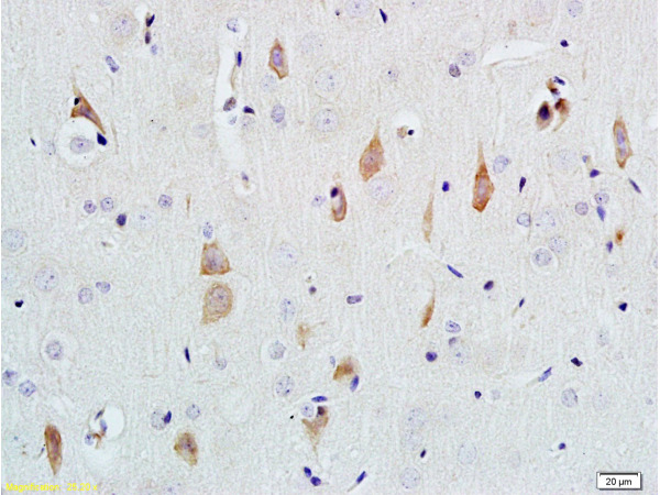
TargetNGF
Overview
- SupplierBoster Bio
- Product NameAnti-NGF Antibody Picoband(r)
- Delivery Days Customer9
- Application Supplier NoteTested Species: In-house tested species with positive results. Other applications have not been tested. Optimal dilutions should be determined by end users.
- ApplicationsWestern Blot
- CertificationResearch Use Only
- ClonalityPolyclonal
- Concentration500 ug/ml
- ConjugateDyLight 488
- Gene ID4803
- Target nameNGF
- Target descriptionnerve growth factor
- Target synonymsBeta-NGF, HSAN5, NGFB, beta-nerve growth factor, nerve growth factor (beta polypeptide), nerve growth factor, beta subunit, pro-nerve growth factor long
- HostRabbit
- IsotypeIgG
- Protein IDP01138
- Protein NameBeta-nerve growth factor
- Scientific DescriptionBoster Bio Anti-NGF Antibody Picoband® catalog # A00341-1. Tested in WB applications. This antibody reacts with Human. The brand Picoband indicates this is a premium antibody that guarantees superior quality, high affinity, and strong signals with minimal background in Western blot applications. Only our best-performing antibodies are designated as Picoband, ensuring unmatched performance.
- ReactivityHuman
- Storage Instruction-20°C,2°C to 8°C
- UNSPSC12352203
Related products
Product group Antibodies
Ngf Polyclonal AntibodyCAC09319
ApplicationsELISA, ImmunoHistoChemistry
TargetNGF
- SizePrice
Product group Antibodies
References
NGFB Polyclonal AntibodyBS-0067R
ApplicationsImmunoFluorescence, Western Blot, ELISA, ImmunoCytoChemistry, ImmunoHistoChemistry, ImmunoHistoChemistry Frozen, ImmunoHistoChemistry Paraffin
ReactivityBovine, Canine, Human, Mouse, Porcine, Rat, Sheep
TargetNGF
- SizePrice
Product group Antibodies
ApplicationsNeutralisation/Blocking
TargetNGF
- SizePrice
Product group Antibodies
Anti-NGF AntibodyA101608
ApplicationsWestern Blot, ELISA
ReactivityHuman
- SizePrice
Product group Antibodies
Anti-NGF Antibody144-00258
ApplicationsWestern Blot
ReactivityHuman, Mouse
TargetNGF
- SizePrice
Product group Antibodies

Anti-Nerve growth factor [mab 911]Ab03301-1.1
ApplicationsELISA, Neutralisation/Blocking, Other Application
ReactivityHuman, Mouse
TargetNGF
- SizePrice
Product group Antibodies
NGF beta antibody [N1C3-2]GTX112043
ApplicationsWestern Blot
ReactivityHuman
TargetNGF
- SizePrice
Product group Antibodies

ApplicationsImmunoPrecipitation, ELISA, Neutralisation/Blocking
ReactivityBovine, Human, Mouse, Reptile
TargetNGF
- SizePrice