


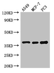
Figure 1. Western blot analysis of SLC9A3R2 using anti-SLC9A3R2 antibody (A05624-2). Electrophoresis was performed on a 5-20% SDS-PAGE gel at 70V (Stacking gel) / 90V (Resolving gel) for 2-3 hours. The sample well of each lane was loaded with 30 ug of sample unde r reducing conditions. Lane 1: human MCF-7 whole cell lysates, Lane 2: human T-47D whole cell lysates, Lane 3: human PC-3 whole cell lysates, Lane 4: mouse lung tissue lysates. After electrophoresis, proteins were transferred to a nitrocellulose membrane at 150 mA for 50-90 minutes. Blocked the membrane with 5% non-fat milk/TBS for 1.5 hour at RT. The membrane was incubated with rabbit anti-SLC9A3R2 antigen affinity purified polyclonal antibody (Catalog # A05624-2) at 0.5 microg/mL overnight at 4°C, then washed with TBS-0.1%Tween 3 times with 5 minutes each and probed with a goat anti-rabbit IgG-HRP secondary antibody at a dilution of 1:5000 for 1.5 hour at RT. The signal is developed using an Enhanced Chemiluminescent detection (ECL) kit (Catalog # EK1002) with Tanon 5200 system. A specific band was detected for SLC9A3R2 at approximately 45 kDa. The expected band size for SLC9A3R2 is at 37 kDa.
. SLC9A3R2 was detected in a paraffin-embedded section of human thyroid cancer tissue. Heat mediated antigen retrieval was performed in EDTA buffer (pH 8.0, epitope retrieval solution). The tissue section was blocked with 10% goat serum. The tissue section was then incubated with 2 microg/ml rabbit anti-SLC9A3R2 Antibody (A05624-2) overnight at 4°C. Peroxidase Conjugated Goat Anti-rabbit IgG was used as secondary antibody and incubated for 30 minutes at 37°C. The tissue section was developed using HRP Conjugated Rabbit IgG Super Vision Assay Kit (Catalog # SV0002) with DAB as the chromogen.")
. SLC9A3R2 was detected in a paraffin-embedded section of human colorectal adenocarcinoma tissue. Heat mediated antigen retrieval was performed in EDTA buffer (pH 8.0, epitope retrieval solution). The tissue section was blocked with 10% goat serum. The tissue section was then incubated with 2 microg/ml rabbit anti-SLC9A3R2 Antibody (A05624-2) overnight at 4°C. Peroxidase Conjugated Goat Anti-rabbit IgG was used as secondary antibody and incubated for 30 minutes at 37°C. The tissue section was developed using HRP Conjugated Rabbit IgG Super Vision Assay Kit (Catalog # SV0002) with DAB as the chromogen.")
. SLC9A3R2 was detected in a paraffin-embedded section of human lung cancer tissue. Heat mediated antigen retrieval was performed in EDTA buffer (pH 8.0, epitope retrieval solution). The tissue section was blocked with 10% goat serum. The tissue section was then incubated with 2 microg/ml rabbit anti-SLC9A3R2 Antibody (A05624-2) overnight at 4°C. Peroxidase Conjugated Goat Anti-rabbit IgG was used as secondary antibody and incubated for 30 minutes at 37°C. The tissue section was developed using HRP Conjugated Rabbit IgG Super Vision Assay Kit (Catalog # SV0002) with DAB as the chromogen.")
. SLC9A3R2 was detected in a paraffin-embedded section of human laryngeal squamous cell carcinoma tissue. Heat mediated antigen retrieval was performed in EDTA buffer (pH 8.0, epitope retrieval solution). The tissue section was blocked with 10% goat serum. The tissue section was then incubated with 2 microg/ml rabbit anti-SLC9A3R2 Antibody (A05624-2) overnight at 4°C. Peroxidase Conjugated Goat Anti-rabbit IgG was used as secondary antibody and incubated for 30 minutes at 37°C. The tissue section was developed using HRP Conjugated Rabbit IgG Super Vision Assay Kit (Catalog # SV0002) with DAB as the chromogen.")
. SLC9A3R2 was detected in a paraffin-embedded section of mouse kidney tissue. Heat mediated antigen retrieval was performed in EDTA buffer (pH 8.0, epitope retrieval solution). The tissue section was blocked with 10% goat serum. The tissue section was then incubated with 2 microg/ml rabbit anti-SLC9A3R2 Antibody (A05624-2) overnight at 4°C. Peroxidase Conjugated Goat Anti-rabbit IgG was used as secondary antibody and incubated for 30 minutes at 37°C. The tissue section was developed using HRP Conjugated Rabbit IgG Super Vision Assay Kit (Catalog # SV0002) with DAB as the chromogen.")
. SLC9A3R2 was detected in a paraffin-embedded section of mouse lung tissue. Heat mediated antigen retrieval was performed in EDTA buffer (pH 8.0, epitope retrieval solution). The tissue section was blocked with 10% goat serum. The tissue section was then incubated with 2 microg/ml rabbit anti-SLC9A3R2 Antibody (A05624-2) overnight at 4°C. Peroxidase Conjugated Goat Anti-rabbit IgG was used as secondary antibody and incubated for 30 minutes at 37°C. The tissue section was developed using HRP Conjugated Rabbit IgG Super Vision Assay Kit (Catalog # SV0002) with DAB as the chromogen.")
. SLC9A3R2 was detected in a paraffin-embedded section of rat kidney tissue. Heat mediated antigen retrieval was performed in EDTA buffer (pH 8.0, epitope retrieval solution). The tissue section was blocked with 10% goat serum. The tissue section was then incubated with 2 microg/ml rabbit anti-SLC9A3R2 Antibody (A05624-2) overnight at 4°C. Peroxidase Conjugated Goat Anti-rabbit IgG was used as secondary antibody and incubated for 30 minutes at 37°C. The tissue section was developed using HRP Conjugated Rabbit IgG Super Vision Assay Kit (Catalog # SV0002) with DAB as the chromogen.")
. SLC9A3R2 was detected in a paraffin-embedded section of rat skin tissue. Heat mediated antigen retrieval was performed in EDTA buffer (pH 8.0, epitope retrieval solution). The tissue section was blocked with 10% goat serum. The tissue section was then incubated with 2 microg/ml rabbit anti-SLC9A3R2 Antibody (A05624-2) overnight at 4°C. Peroxidase Conjugated Goat Anti-rabbit IgG was used as secondary antibody and incubated for 30 minutes at 37°C. The tissue section was developed using HRP Conjugated Rabbit IgG Super Vision Assay Kit (Catalog # SV0002) with DAB as the chromogen.")
. SLC9A3R2 was detected in a paraffin-embedded section of rat lung tissue. Heat mediated antigen retrieval was performed in EDTA buffer (pH 8.0, epitope retrieval solution). The tissue section was blocked with 10% goat serum. The tissue section was then incubated with 2 microg/ml rabbit anti-SLC9A3R2 Antibody (A05624-2) overnight at 4°C. Peroxidase Conjugated Goat Anti-rabbit IgG was used as secondary antibody and incubated for 30 minutes at 37°C. The tissue section was developed using HRP Conjugated Rabbit IgG Super Vision Assay Kit (Catalog # SV0002) with DAB as the chromogen.")
Figure 1. Western blot analysis of SLC9A3R2 using anti-SLC9A3R2 antibody (A05624-2). Electrophoresis was performed on a 5-20% SDS-PAGE gel at 70V (Stacking gel) / 90V (Resolving gel) for 2-3 hours. The sample well of each lane was loaded with 30 ug of sample unde r reducing conditions. Lane 1: human MCF-7 whole cell lysates, Lane 2: human T-47D whole cell lysates, Lane 3: human PC-3 whole cell lysates, Lane 4: mouse lung tissue lysates. After electrophoresis, proteins were transferred to a nitrocellulose membrane at 150 mA for 50-90 minutes. Blocked the membrane with 5% non-fat milk/TBS for 1.5 hour at RT. The membrane was incubated with rabbit anti-SLC9A3R2 antigen affinity purified polyclonal antibody (Catalog # A05624-2) at 0.5 microg/mL overnight at 4°C, then washed with TBS-0.1%Tween 3 times with 5 minutes each and probed with a goat anti-rabbit IgG-HRP secondary antibody at a dilution of 1:5000 for 1.5 hour at RT. The signal is developed using an Enhanced Chemiluminescent detection (ECL) kit (Catalog # EK1002) with Tanon 5200 system. A specific band was detected for SLC9A3R2 at approximately 45 kDa. The expected band size for SLC9A3R2 is at 37 kDa.
Anti-NHERF-2/SIP-1/SLC9A3R2 Antibody Picoband(r)
A05624-2-CARRIER-FREE
ApplicationsFlow Cytometry, Western Blot, ELISA, ImmunoHistoChemistry
Product group Antibodies
ReactivityHuman, Mouse, Rat
TargetNHERF2
Overview
- SupplierBoster Bio
- Product NameAnti-NHERF-2/SIP-1/SLC9A3R2 Antibody Picoband(r)
- Delivery Days Customer9
- ApplicationsFlow Cytometry, Western Blot, ELISA, ImmunoHistoChemistry
- CertificationResearch Use Only
- ClonalityPolyclonal
- Concentration500 ug/ml
- Gene ID9351
- Target nameNHERF2
- Target descriptionNHERF family PDZ scaffold protein 2
- Target synonymsE3KARP, NHE3RF2, NHERF-2, OCTS2, SIP-1, SIP1, SLC9A3R2, TKA-1, Na(+)/H(+) exchange regulatory cofactor NHE-RF2, NHE3 kinase A regulatory protein E3KARP, NHE3 regulatory factor 2, Na(+)/H(+) exchange regulatory cofactor 2, SLC9A3 regulator 2, SRY-interacting protein 1, sodium/hydrogen exchanger 3 kinase A regulatory protein, solute carrier family 9 (sodium/hydrogen exchanger), member 3 regulator 2, solute carrier family 9, subfamily A (NHE3, cation proton antiporter 3), member 3 regulator 2, tyrosine kinase activator protein 1
- HostRabbit
- IsotypeIgG
- Protein IDQ15599
- Protein NameNa(+)/H(+) exchange regulatory cofactor NHE-RF2
- Scientific DescriptionBoster Bio Anti-NHERF-2/SIP-1/SLC9A3R2 Antibody Picoband® catalog # A05624-2. Tested in ELISA, Flow Cytometry, IHC, WB applications. This antibody reacts with Human, Mouse, Rat. The brand Picoband indicates this is a premium antibody that guarantees superior quality, high affinity, and strong signals with minimal background in Western blot applications. Only our best-performing antibodies are designated as Picoband, ensuring unmatched performance.
- ReactivityHuman, Mouse, Rat
- Storage Instruction-20°C,2°C to 8°C
- UNSPSC12352203
Related products
Product group Antibodies
SLC9A3R2 AntibodyCSB-PA01759A0RB
ApplicationsWestern Blot, ELISA, ImmunoHistoChemistry
ReactivityHuman
TargetNHERF2
- SizePrice
Product group Antibodies
Anti-SLC9A3R2 Antibody144-65015
ApplicationsWestern Blot
ReactivityHuman
TargetNHERF2
- SizePrice
Product group Antibodies
ApplicationsWestern Blot, ELISA
ReactivityHuman
- SizePrice
Product group Antibodies

ApplicationsWestern Blot, ELISA
ReactivityHuman
TargetNHERF2
- SizePrice

Product group Antibodies
Anti-SLC9A3R2 AntibodyHPA001672
ApplicationsImmunoHistoChemistry
ReactivityHuman
TargetNHERF2
- SizePrice
Product group Antibodies

SLC9A3R2 / SIP1 AntibodyLS-C405799
ApplicationsELISA, ImmunoHistoChemistry
ReactivityHuman, Mouse, Rat
TargetNHERF2
- SizePrice
Product group Antibodies
NHERF2 antibody [N2C3]GTX101466
ApplicationsWestern Blot
ReactivityHuman, Mouse
TargetNHERF2
- SizePrice
Product group Antibodies
SLC9A3R2 Polyclonal AntibodyCAC14689
ApplicationsWestern Blot, ELISA, ImmunoHistoChemistry
TargetNHERF2
- SizePrice