


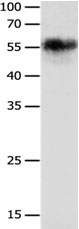
Figure 1. Western blot analysis of CHRNA3 using anti-CHRNA3 antibody (A01981-1). Electrophoresis was performed on a 5-20% SDS-PAGE gel at 70V (Stacking gel) / 90V (Resolving gel) for 2-3 hours. The sample well of each lane was loaded with 50ug of sample under reducing conditions. Lane 1: human Hela whole cell lysates, Lane 2: human MDA-MB-453 whole cell lysates, Lane 3: human Jurkat whole cell lysates, Lane 4: human HepG2 whole cell lysates, Lane 5: human SK-OV-3 whole cell lysates, Lane 6: human PANC-1 whole cell lysates, Lane 7: mouse thymus tissue lysates. After Electrophoresis, proteins were transferred to a Nitrocellulose membrane at 150mA for 50-90 minutes. Blocked the membrane with 5% Non-fat Milk/ TBS for 1.5 hour at RT. The membrane was incubated with rabbit anti-CHRNA3 antigen affinity purified polyclonal antibody (Catalog # A01981-1) at 0.5 microg/mL overnight at 4°C, then washed with TBS-0.1%Tween 3 times with 5 minutes each and probed with a goat anti-rabbit IgG-HRP secondary antibody at a dilution of 1:10000 for 1.5 hour at RT. The signal is developed using an Enhanced Chemiluminescent detection (ECL) kit (Catalog # EK1002) with Tanon 5200 system. A specific band was detected for CHRNA3 at approximately 60KD. The expected band size for CHRNA3 is at 57KD.
. Overlay histogram showing U251 cells stained with A01981-1 (Blue line). To facilitate intracellular staining, cells were fixed with 4% paraformaldehyde and permeabilized with permeabilization buffer. The cells were blocked with 10% normal goat serum. And then incubated with rabbit anti-CHRNA3 Antibody (A01981-1,1microg/1x106 cells) for 30 min at 20°C. DyLight®488 conjugated goat anti-rabbit IgG (BA1127, 5-10microg/1x106 cells) was used as secondary antibody for 30 minutes at 20°C. Isotype control antibody (Green line) was rabbit IgG (1microg/1x106) used under the same conditions. Unlabelled sample without incubation with primary antibody and secondary antibody (Red line) was used as a blank control.")
. Overlay histogram showing U-87 cells stained with A01981-1 (Blue line). To facilitate intracellular staining, cells were fixed with 4% paraformaldehyde and permeabilized with permeabilization buffer. The cells were blocked with 10% normal goat serum. And then incubated with rabbit anti-CHRNA3 Antibody (A01981-1,1microg/1x106 cells) for 30 min at 20°C. DyLight®488 conjugated goat anti-rabbit IgG (BA1127, 5-10microg/1x106 cells) was used as secondary antibody for 30 minutes at 20°C. Isotype control antibody (Green line) was rabbit IgG (1microg/1x106) used under the same conditions. Unlabelled sample without incubation with primary antibody and secondary antibody (Red line) was used as a blank control.")
Figure 1. Western blot analysis of CHRNA3 using anti-CHRNA3 antibody (A01981-1). Electrophoresis was performed on a 5-20% SDS-PAGE gel at 70V (Stacking gel) / 90V (Resolving gel) for 2-3 hours. The sample well of each lane was loaded with 50ug of sample under reducing conditions. Lane 1: human Hela whole cell lysates, Lane 2: human MDA-MB-453 whole cell lysates, Lane 3: human Jurkat whole cell lysates, Lane 4: human HepG2 whole cell lysates, Lane 5: human SK-OV-3 whole cell lysates, Lane 6: human PANC-1 whole cell lysates, Lane 7: mouse thymus tissue lysates. After Electrophoresis, proteins were transferred to a Nitrocellulose membrane at 150mA for 50-90 minutes. Blocked the membrane with 5% Non-fat Milk/ TBS for 1.5 hour at RT. The membrane was incubated with rabbit anti-CHRNA3 antigen affinity purified polyclonal antibody (Catalog # A01981-1) at 0.5 microg/mL overnight at 4°C, then washed with TBS-0.1%Tween 3 times with 5 minutes each and probed with a goat anti-rabbit IgG-HRP secondary antibody at a dilution of 1:10000 for 1.5 hour at RT. The signal is developed using an Enhanced Chemiluminescent detection (ECL) kit (Catalog # EK1002) with Tanon 5200 system. A specific band was detected for CHRNA3 at approximately 60KD. The expected band size for CHRNA3 is at 57KD.
Anti-Nicotinic Acetylcholine Receptor alpha 3/CHRNA3 Antibody Picoband(r)
A01981-1-CARRIER-FREE
ApplicationsFlow Cytometry, Western Blot
Product group Antibodies
ReactivityHuman, Mouse, Rat
TargetCHRNA3
Overview
- SupplierBoster Bio
- Product NameAnti-Nicotinic Acetylcholine Receptor alpha 3/CHRNA3 Antibody Picoband(r)
- Delivery Days Customer9
- ApplicationsFlow Cytometry, Western Blot
- CertificationResearch Use Only
- ClonalityPolyclonal
- Concentration500 ug/ml
- Gene ID1136
- Target nameCHRNA3
- Target descriptioncholinergic receptor nicotinic alpha 3 subunit
- Target synonymsBAIPRCK, LNCR2, NACHRA3, PAOD2, neuronal acetylcholine receptor subunit alpha-3, cholinergic receptor, nicotinic alpha 3, cholinergic receptor, nicotinic, alpha 3 (neuronal), cholinergic receptor, nicotinic, alpha polypeptide 3, neuronal nicotinic acetylcholine receptor, alpha3 subunit
- HostRabbit
- IsotypeIgG
- Protein IDP32297
- Protein NameNeuronal acetylcholine receptor subunit alpha-3
- Scientific DescriptionBoster Bio Anti-Nicotinic Acetylcholine Receptor alpha 3/CHRNA3 Antibody Picoband® catalog # A01981-1. Tested in Flow Cytometry, WB applications. This antibody reacts with Human, Mouse, Rat. The brand Picoband indicates this is a premium antibody that guarantees superior quality, high affinity, and strong signals with minimal background in Western blot applications. Only our best-performing antibodies are designated as Picoband, ensuring unmatched performance.
- ReactivityHuman, Mouse, Rat
- Storage Instruction-20°C,2°C to 8°C
- UNSPSC12352203
Related products
Product group Antibodies
CHRNA3 AntibodyCSB-PA085715
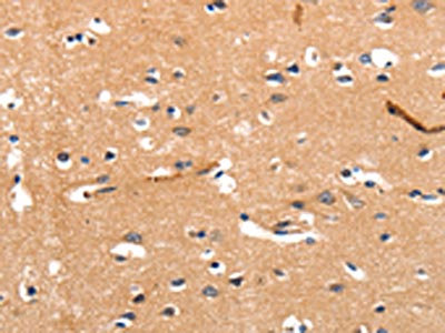
ApplicationsWestern Blot, ELISA, ImmunoHistoChemistry
ReactivityHuman, Mouse, Rat
TargetCHRNA3
- SizePrice
Product group Antibodies
Anti-CHRNA3 AntibodyA47690
ApplicationsWestern Blot, ELISA, ImmunoHistoChemistry
ReactivityHuman, Mouse, Rat
- SizePrice
Product group Antibodies

CHRNA3 AntibodyLS-C765529
ApplicationsWestern Blot, ELISA, ImmunoHistoChemistry
ReactivityHuman, Mouse, Rat
TargetCHRNA3
- SizePrice
Product group Antibodies
ApplicationsImmunoPrecipitation, Western Blot, ImmunoCytoChemistry, ImmunoHistoChemistry
ReactivityMouse, Porcine, Rat
TargetCHRNA3
- SizePrice
Product group Antibodies
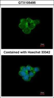
AChR alpha 3 antibodyGTX105495
ApplicationsImmunoFluorescence, Western Blot, ImmunoCytoChemistry
ReactivityHuman
TargetCHRNA3
- SizePrice
Product group Antibodies
CHRNA3 Polyclonal AntibodyBS-6455R
ApplicationsImmunoFluorescence, Western Blot, ELISA, ImmunoCytoChemistry, ImmunoHistoChemistry, ImmunoHistoChemistry Frozen, ImmunoHistoChemistry Paraffin
ReactivityBovine, Canine, Chicken, Equine, Human, Mouse, Porcine, Rabbit, Rat, Sheep
TargetCHRNA3
- SizePrice