


Anti-NMDAR1 Antibody
A83429
ApplicationsImmunoFluorescence, ELISA
Product group Antibodies
ReactivityHuman, Mouse
Overview
- SupplierAntibodies.com
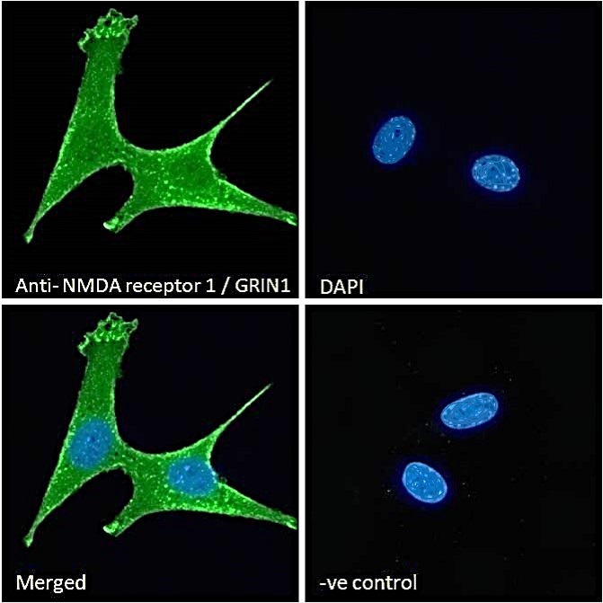
- Product NameAnti-NMDAR1 Antibody
- Delivery Days Customer7
- ApplicationsImmunoFluorescence, ELISA
- CertificationResearch Use Only
- ClonalityPolyclonal
- Concentration500 ug/ml
- ConjugateUnconjugated
- HostGoat
- IsotypeIgG
- Scientific DescriptionGoat polyclonal antibody to NMDAR1.
- ReactivityHuman, Mouse
- UNSPSC12352203
Related products
Product group Antibodies
Anti-GRIN1 Antibody144-66462
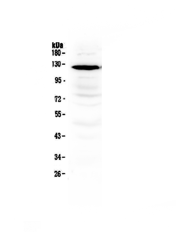
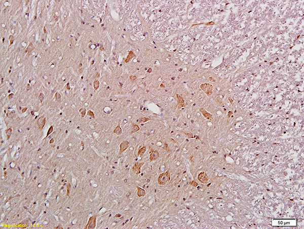
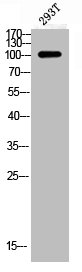
ApplicationsImmunoFluorescence, Western Blot, ImmunoHistoChemistry
ReactivityHuman, Mouse, Rat
TargetGRIN1
- SizePrice
Product group Antibodies
Anti-NMDAR1/GRIN1 Antibody Picoband(r)A01808-CARRIER-FREE
ApplicationsWestern Blot
ReactivityHuman, Mouse, Rat
TargetGRIN1
- SizePrice
Product group Antibodies
References
NMDAR1 Polyclonal AntibodyBS-2175R
ApplicationsImmunoFluorescence, Western Blot, ELISA, ImmunoCytoChemistry, ImmunoHistoChemistry, ImmunoHistoChemistry Frozen, ImmunoHistoChemistry Paraffin
ReactivityCanine, Chicken, Human, Mouse, Porcine, Rabbit, Rat
TargetGRIN1
- SizePrice
Product group Antibodies
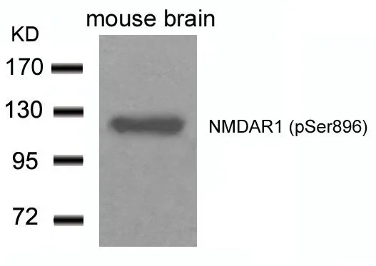
Phospho-GRIN1 (S896) AntibodyCSB-PA008967
ApplicationsWestern Blot, ELISA, ImmunoHistoChemistry
ReactivityHuman, Mouse, Rat
TargetGRIN1
- SizePrice
Product group Antibodies

ApplicationsImmunoFluorescence, ELISA
ReactivityCanine, Human, Mouse, Rat
TargetGRIN1
- SizePrice
Product group Antibodies
Grin1 Recombinant AntibodyCAC12218
ApplicationsFlow Cytometry, ELISA
TargetGRIN1
- SizePrice
Product group Antibodies

GRIN1 / NMDAR1 AntibodyLS-C402488
ApplicationsELISA, ImmunoHistoChemistry
ReactivityHuman, Mouse, Rat
TargetGRIN1
- SizePrice

Product group Antibodies
Anti-GRIN1 AntibodyHPA067773
ApplicationsImmunoHistoChemistry
ReactivityHuman
TargetGRIN1
- SizePrice
Product group Antibodies
NMDAR1 (phospho Ser896) antibodyGTX50168
ApplicationsWestern Blot
ReactivityHuman, Mouse
TargetGRIN1
- SizePrice