


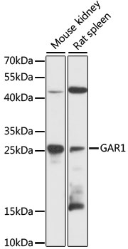
Figure 1. Western blot analysis of NOLA1/GAR1 using anti-NOLA1/GAR1 antibody (A07049-1). Electrophoresis was performed on a 5-20% SDS-PAGE gel at 70V (Stacking gel) / 90V (Resolving gel) for 2-3 hours. The sample well of each lane was loaded with 30 ug of sample under reducing conditions. Lane 1: human A375 whole cell lysates, Lane 2: human K562 whole cell lysates, Lane 3: human HL-60 whole cell lysates, Lane 4: human MCF-7 whole cell lysates. After electrophoresis, proteins were transferred to a nitrocellulose membrane at 150 mA for 50-90 minutes. Blocked the membrane with 5% non-fat milk/TBS for 1.5 hour at RT. The membrane was incubated with rabbit anti-NOLA1/GAR1 antigen affinity purified polyclonal antibody (Catalog # A07049-1) at 0.5 microg/mL overnight at 4°C, then washed with TBS-0.1%Tween 3 times with 5 minutes each and probed with a goat anti-rabbit IgG-HRP secondary antibody at a dilution of 1:5000 for 1.5 hour at RT. The signal is developed using an Enhanced Chemiluminescent detection (ECL) kit (Catalog # EK1002) with Tanon 5200 system. A specific band was detected for NOLA1/GAR1 at approximately 25 kDa. The expected band size for NOLA1/GAR1 is at 25 kDa.
. NOLA1/GAR1 was detected in a paraffin-embedded section of human breast cancer tissue. Heat mediated antigen retrieval was performed in EDTA buffer (pH 8.0, epitope retrieval solution). The tissue section was blocked with 10% goat serum. The tissue section was then incubated with 2 microg/ml rabbit anti-NOLA1/GAR1 Antibody (A07049-1) overnight at 4°C. Biotinylated goat anti-rabbit IgG was used as secondary antibody and incubated for 30 minutes at 37°C. The tissue section was developed using Strepavidin-Biotin-Complex (SABC) (Catalog # SA1022) with DAB as the chromogen.")
. NOLA1/GAR1 was detected in a paraffin-embedded section of human gastric carcinoma tissue. Heat mediated antigen retrieval was performed in EDTA buffer (pH 8.0, epitope retrieval solution). The tissue section was blocked with 10% goat serum. The tissue section was then incubated with 2 microg/ml rabbit anti-NOLA1/GAR1 Antibody (A07049-1) overnight at 4°C. Biotinylated goat anti-rabbit IgG was used as secondary antibody and incubated for 30 minutes at 37°C. The tissue section was developed using Strepavidin-Biotin-Complex (SABC) (Catalog # SA1022) with DAB as the chromogen.")
. NOLA1/GAR1 was detected in a paraffin-embedded section of human liver cancer tissue. Heat mediated antigen retrieval was performed in EDTA buffer (pH 8.0, epitope retrieval solution). The tissue section was blocked with 10% goat serum. The tissue section was then incubated with 2 microg/ml rabbit anti-NOLA1/GAR1 Antibody (A07049-1) overnight at 4°C. Biotinylated goat anti-rabbit IgG was used as secondary antibody and incubated for 30 minutes at 37°C. The tissue section was developed using Strepavidin-Biotin-Complex (SABC) (Catalog # SA1022) with DAB as the chromogen.")
. NOLA1/GAR1 was detected in a paraffin-embedded section of human ovarian adenoma tissue. Heat mediated antigen retrieval was performed in EDTA buffer (pH 8.0, epitope retrieval solution). The tissue section was blocked with 10% goat serum. The tissue section was then incubated with 2 microg/ml rabbit anti-NOLA1/GAR1 Antibody (A07049-1) overnight at 4°C. Biotinylated goat anti-rabbit IgG was used as secondary antibody and incubated for 30 minutes at 37°C. The tissue section was developed using Strepavidin-Biotin-Complex (SABC) (Catalog # SA1022) with DAB as the chromogen.")
. NOLA1/GAR1 was detected in a paraffin-embedded section of human pancreatic cancer tissue. Heat mediated antigen retrieval was performed in EDTA buffer (pH 8.0, epitope retrieval solution). The tissue section was blocked with 10% goat serum. The tissue section was then incubated with 2 microg/ml rabbit anti-NOLA1/GAR1 Antibody (A07049-1) overnight at 4°C. Biotinylated goat anti-rabbit IgG was used as secondary antibody and incubated for 30 minutes at 37°C. The tissue section was developed using Strepavidin-Biotin-Complex (SABC) (Catalog # SA1022) with DAB as the chromogen.")
. NOLA1/GAR1 was detected in a paraffin-embedded section of human prostate cancer tissue. Heat mediated antigen retrieval was performed in EDTA buffer (pH 8.0, epitope retrieval solution). The tissue section was blocked with 10% goat serum. The tissue section was then incubated with 2 microg/ml rabbit anti-NOLA1/GAR1 Antibody (A07049-1) overnight at 4°C. Biotinylated goat anti-rabbit IgG was used as secondary antibody and incubated for 30 minutes at 37°C. The tissue section was developed using Strepavidin-Biotin-Complex (SABC) (Catalog # SA1022) with DAB as the chromogen.")
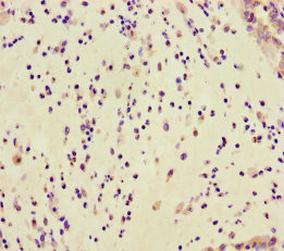
. NOLA1/GAR1 was detected in a paraffin-embedded section of human glioma tissue. Heat mediated antigen retrieval was performed in EDTA buffer (pH 8.0, epitope retrieval solution). The tissue section was blocked with 10% goat serum. The tissue section was then incubated with 2 microg/ml rabbit anti-NOLA1/GAR1 Antibody (A07049-1) overnight at 4°C. Biotinylated goat anti-rabbit IgG was used as secondary antibody and incubated for 30 minutes at 37°C. The tissue section was developed using Strepavidin-Biotin-Complex (SABC) (Catalog # SA1022) with DAB as the chromogen.")
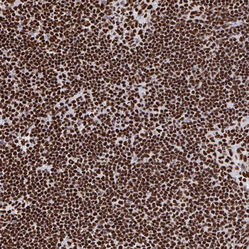
. NOLA1/GAR1 was detected in a paraffin-embedded section of human lymphoma tissue. Heat mediated antigen retrieval was performed in EDTA buffer (pH 8.0, epitope retrieval solution). The tissue section was blocked with 10% goat serum. The tissue section was then incubated with 2 microg/ml rabbit anti-NOLA1/GAR1 Antibody (A07049-1) overnight at 4°C. Biotinylated goat anti-rabbit IgG was used as secondary antibody and incubated for 30 minutes at 37°C. The tissue section was developed using Strepavidin-Biotin-Complex (SABC) (Catalog # SA1022) with DAB as the chromogen.")
. NOLA1/GAR1 was detected in an immunocytochemical section of MCF-7 cells. Enzyme antigen retrieval was performed using IHC enzyme antigen retrieval reagent (AR0022) for 15 mins. The cells were blocked with 10% goat serum. And then incubated with 5 microg/mL rabbit anti-NOLA1/GAR1 Antibody (A07049-1) overnight at 4°C. DyLight®488 Conjugated Goat Anti-Rabbit IgG (BA1127) was used as secondary antibody at 1:100 dilution and incubated for 30 minutes at 37°C. The section was counterstained with DAPI. Visualize using a fluorescence microscope and filter sets appropriate for the label used.")
Figure 1. Western blot analysis of NOLA1/GAR1 using anti-NOLA1/GAR1 antibody (A07049-1). Electrophoresis was performed on a 5-20% SDS-PAGE gel at 70V (Stacking gel) / 90V (Resolving gel) for 2-3 hours. The sample well of each lane was loaded with 30 ug of sample under reducing conditions. Lane 1: human A375 whole cell lysates, Lane 2: human K562 whole cell lysates, Lane 3: human HL-60 whole cell lysates, Lane 4: human MCF-7 whole cell lysates. After electrophoresis, proteins were transferred to a nitrocellulose membrane at 150 mA for 50-90 minutes. Blocked the membrane with 5% non-fat milk/TBS for 1.5 hour at RT. The membrane was incubated with rabbit anti-NOLA1/GAR1 antigen affinity purified polyclonal antibody (Catalog # A07049-1) at 0.5 microg/mL overnight at 4°C, then washed with TBS-0.1%Tween 3 times with 5 minutes each and probed with a goat anti-rabbit IgG-HRP secondary antibody at a dilution of 1:5000 for 1.5 hour at RT. The signal is developed using an Enhanced Chemiluminescent detection (ECL) kit (Catalog # EK1002) with Tanon 5200 system. A specific band was detected for NOLA1/GAR1 at approximately 25 kDa. The expected band size for NOLA1/GAR1 is at 25 kDa.
Anti-NOLA1/GAR1 Antibody Picoband(r)
A07049-1-HRP
ApplicationsFlow Cytometry, ImmunoFluorescence, Western Blot, ELISA, ImmunoCytoChemistry, ImmunoHistoChemistry
Product group Antibodies
ReactivityHuman
TargetGAR1
Overview
- SupplierBoster Bio
- Product NameAnti-NOLA1/GAR1 Antibody Picoband(r)
- Delivery Days Customer9
- ApplicationsFlow Cytometry, ImmunoFluorescence, Western Blot, ELISA, ImmunoCytoChemistry, ImmunoHistoChemistry
- CertificationResearch Use Only
- ClonalityPolyclonal
- Concentration500 ug/ml
- ConjugateHRP
- Gene ID54433
- Target nameGAR1
- Target descriptionGAR1 ribonucleoprotein
- Target synonymsNOLA1, H/ACA ribonucleoprotein complex subunit 1, GAR1 homolog, ribonucleoprotein, GAR1 ribonucleoprotein homolog, nucleolar protein family A member 1, nucleolar protein family A, member 1 (H/ACA small nucleolar RNPs), snoRNP protein GAR1
- HostRabbit
- IsotypeIgG
- Protein IDQ9NY12
- Protein NameH/ACA ribonucleoprotein complex subunit 1
- Scientific DescriptionBoster Bio Anti-NOLA1/GAR1 Antibody Picoband® catalog # A07049-1. Tested in ELISA, Flow Cytometry, IF, IHC, ICC, WB applications. This antibody reacts with Human. The brand Picoband indicates this is a premium antibody that guarantees superior quality, high affinity, and strong signals with minimal background in Western blot applications. Only our best-performing antibodies are designated as Picoband, ensuring unmatched performance.
- ReactivityHuman
- Storage Instruction-20°C,2°C to 8°C
- UNSPSC12352203
Related products
Product group Antibodies
Anti-GAR1 (Center) Antibody102-23597
ApplicationsWestern Blot
TargetGAR1
- SizePrice
Product group Antibodies
Anti-NOLA1 AntibodyA89082
ApplicationsWestern Blot
ReactivityMouse, Rat
- SizePrice
Product group Antibodies

GAR1 / NOLA1 AntibodyLS-C747826
ApplicationsWestern Blot
ReactivityHuman, Mouse, Rat
TargetGAR1
- SizePrice
Product group Antibodies
Anti-GAR1 AntibodyHPA059098
ApplicationsImmunoCytoChemistry, ImmunoHistoChemistry
ReactivityHuman
TargetGAR1
- SizePrice
Product group Antibodies
GAR1 AntibodyCSB-PA878917LA01HU
ApplicationsELISA, ImmunoHistoChemistry
ReactivityHuman
TargetGAR1
- SizePrice
Product group Antibodies
Anti-NOLA1/GAR1 Antibody Picoband(r)A07049-1-CARRIER-FREE
ApplicationsFlow Cytometry, ImmunoFluorescence, Western Blot, ELISA, ImmunoCytoChemistry, ImmunoHistoChemistry
ReactivityHuman
TargetGAR1
- SizePrice