

Anti-NOS1 Antibody
A101356
ApplicationsWestern Blot, ELISA
Product group Antibodies
ReactivityHuman
Overview
- SupplierAntibodies.com
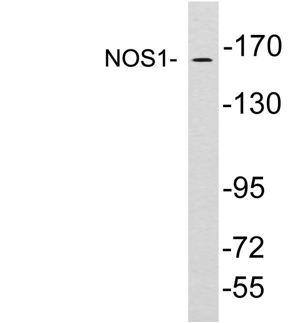
- Product NameAnti-NOS1 Antibody
- Delivery Days Customer7
- ApplicationsWestern Blot, ELISA
- CertificationResearch Use Only
- ClonalityPolyclonal
- ConjugateUnconjugated
- HostRabbit
- IsotypeIgG
- Scientific DescriptionRabbit polyclonal antibody to NOS1.
- ReactivityHuman
- UNSPSC12352203
Related products

Product group Antibodies
Anti-nNOS (neuronal)/NOS1 Antibody Picoband(r)A01070-2-CARRIER-FREE
ApplicationsFlow Cytometry, Western Blot, ELISA
ReactivityHuman, Mouse, Rat
TargetNOS1
- SizePrice
Product group Antibodies
Anti-Rat NOS1 Antibody144-01485
ApplicationsWestern Blot
ReactivityHuman, Rat
TargetNOS1
- SizePrice
Product group Antibodies

NOS1 / nNOS AntibodyLS-C760906
ApplicationsWestern Blot
ReactivityHuman, Mouse, Rat
TargetNOS1
- SizePrice
Product group Antibodies
References
nNos Polyclonal AntibodyBS-0156R
ApplicationsImmunoFluorescence, Western Blot, ELISA, ImmunoCytoChemistry, ImmunoHistoChemistry, ImmunoHistoChemistry Frozen, ImmunoHistoChemistry Paraffin
ReactivityHuman, Mouse, Rabbit, Rat
TargetNOS1
- SizePrice
Product group Antibodies
NOS1 AntibodyCSB-PA003463
ApplicationsImmunoFluorescence, Western Blot, ELISA, ImmunoHistoChemistry
ReactivityHuman, Mouse, Rat
TargetNOS1
- SizePrice
Product group Antibodies
References

Goat anti-NOS1EB05259
ApplicationsFlow Cytometry, ImmunoFluorescence, ELISA, ImmunoHistoChemistry
ReactivityBovine, Canine, Human, Mouse, Rat
TargetNOS1
- SizePrice
Product group Antibodies
NOS1 Polyclonal AntibodyCAC14656
ApplicationsImmunoFluorescence, Western Blot, ELISA
ReactivityMouse
TargetNOS1
- SizePrice
Product group Antibodies
nNOS antibodyGTX132857
ApplicationsWestern Blot
ReactivityHuman, Mouse, Rat
TargetNOS1
- SizePrice

Product group Antibodies
Anti-NOS1 AntibodyHPA058312
ApplicationsImmunoCytoChemistry
ReactivityHuman
TargetNOS1
- SizePrice