


Immunohistochemical staining of human testis shows strong granular cytoplasmic positivity in cells in seminiferous ducts and Leydig cells.
 shows similar pattern to independent antibody HPA023295 (B).")
Immunohistochemical staining of human testis shows strong granular cytoplasmic positivity in cells in seminiferous ducts and Leydig cells.
Anti-NPLOC4 Antibody
HPA021560
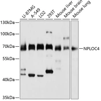
ApplicationsWestern Blot, ImmunoCytoChemistry, ImmunoHistoChemistry
Product group Antibodies
ReactivityHuman, Mouse, Rat
TargetNPLOC4
Overview
- SupplierAtlas Antibodies
- Product NameAnti-NPLOC4 Antibody
- Delivery Days Customer4
- ApplicationsWestern Blot, ImmunoCytoChemistry, ImmunoHistoChemistry
- CertificationResearch Use Only
- ClonalityPolyclonal
- ConjugateUnconjugated
- Gene ID55666
- Target nameNPLOC4
- Target descriptionNPL4 homolog, ubiquitin recognition factor
- Target synonymsNPL4, nuclear protein localization protein 4 homolog, NPLOC4 ubiquitin recognition factor, nuclear protein localization 4 homolog
- HostRabbit
- IsotypeIgG
- Protein IDQ8TAT6
- Protein NameNuclear protein localization protein 4 homolog
- Scientific DescriptionRecombinant Protein Epitope Signature Tag (PrEST) antigen sequence
- ReactivityHuman, Mouse, Rat
- Storage Instruction-20°C,2°C to 8°C
- UNSPSC41116161
Datasheet
MSDS
Related products
Product group Antibodies
Anti-NPLOC4 Antibody144-03256
ApplicationsWestern Blot
ReactivityHuman, Mouse, Rat
TargetNPLOC4
- SizePrice
Product group Antibodies
NPL4 antibodyGTX66423
ApplicationsImmunoFluorescence, Western Blot, ImmunoCytoChemistry
ReactivityHuman, Mouse
TargetNPLOC4
- SizePrice
Product group Antibodies
NPLOC4 AntibodyCSB-PA015995GA01HU
ApplicationsWestern Blot, ELISA
ReactivityHuman, Mouse, Rat
TargetNPLOC4
- SizePrice
Product group Antibodies

NPLOC4 AntibodyLS-C497961
ApplicationsWestern Blot
ReactivityHuman, Mouse, Rat
TargetNPLOC4
- SizePrice

Product group Antibodies
Anti-NPLOC4 AntibodyHPA023295
ApplicationsWestern Blot, ImmunoCytoChemistry, ImmunoHistoChemistry
ReactivityHuman
TargetNPLOC4
- SizePrice

Product group Antibodies
Anti-NPL4/NPLOC4 Antibody Picoband(r)A08476-1-CARRIER-FREE
ApplicationsImmunoFluorescence, Western Blot, ELISA, ImmunoCytoChemistry
ReactivityHuman, Mouse, Rat
TargetNPLOC4
- SizePrice