


Figure 1. Western blot analysis of KDM1B using anti-KDM1B antibody (M01077-1). Electrophoresis was performed on a 5-20% SDS-PAGE gel at 70V (Stacking gel) / 90V (Resolving gel) for 2-3 hours. The sample well of each lane was loaded with 30 ug of sample under reducing conditions. Lane 1: human Hela whole cell lysates, Lane 2: human HepG2 whole cell lysates, Lane 3: human U-87MG whole cell lysates, Lane 4: human U2OS whole cell lysates, Lane 5: rat liver tissue lysates, Lane 6: rat RH35 whole cell lysates, Lane 7: mouse liver tissue lysates, Lane 8: mouse HEPA1-6 whole cell lysates. After electrophoresis, proteins were transferred to a nitrocellulose membrane at 150 mA for 50-90 minutes. Blocked the membrane with 5% non-fat milk/TBS for 1.5 hour at RT. The membrane was incubated with rabbit anti-KDM1B antigen affinity purified monoclonal antibody (Catalog # M01077-1) at 1:500 overnight at 4°C, then washed with TBS-0.1%Tween 3 times with 5 minutes each and probed with a goat anti-rabbit IgG-HRP secondary antibody at a dilution of 1:500 for 1.5 hour at RT. The signal is developed using an Enhanced Chemiluminescent detection (ECL) kit (Catalog # EK1002) with Tanon 5200 system. A specific band was detected for KDM1B at approximately 55 kDa. The expected band size for KDM1B is at 67 kDa.
Figure 1. Western blot analysis of KDM1B using anti-KDM1B antibody (M01077-1). Electrophoresis was performed on a 5-20% SDS-PAGE gel at 70V (Stacking gel) / 90V (Resolving gel) for 2-3 hours. The sample well of each lane was loaded with 30 ug of sample under reducing conditions. Lane 1: human Hela whole cell lysates, Lane 2: human HepG2 whole cell lysates, Lane 3: human U-87MG whole cell lysates, Lane 4: human U2OS whole cell lysates, Lane 5: rat liver tissue lysates, Lane 6: rat RH35 whole cell lysates, Lane 7: mouse liver tissue lysates, Lane 8: mouse HEPA1-6 whole cell lysates. After electrophoresis, proteins were transferred to a nitrocellulose membrane at 150 mA for 50-90 minutes. Blocked the membrane with 5% non-fat milk/TBS for 1.5 hour at RT. The membrane was incubated with rabbit anti-KDM1B antigen affinity purified monoclonal antibody (Catalog # M01077-1) at 1:500 overnight at 4°C, then washed with TBS-0.1%Tween 3 times with 5 minutes each and probed with a goat anti-rabbit IgG-HRP secondary antibody at a dilution of 1:500 for 1.5 hour at RT. The signal is developed using an Enhanced Chemiluminescent detection (ECL) kit (Catalog # EK1002) with Tanon 5200 system. A specific band was detected for KDM1B at approximately 55 kDa. The expected band size for KDM1B is at 67 kDa.
Anti-NR1D1 Rabbit Monoclonal Antibody
M01077-1
ApplicationsFlow Cytometry, ImmunoFluorescence, Western Blot, ImmunoCytoChemistry, ImmunoHistoChemistry
Product group Antibodies
ReactivityHuman, Mouse, Rat
TargetNR1D1
Overview
- SupplierBoster Bio
- Product NameAnti-NR1D1 Rabbit Monoclonal Antibody
- Delivery Days Customer9
- ApplicationsFlow Cytometry, ImmunoFluorescence, Western Blot, ImmunoCytoChemistry, ImmunoHistoChemistry
- CertificationResearch Use Only
- ClonalityMonoclonal
- Clone ID18N41
- Gene ID9572
- Target nameNR1D1
- Target descriptionnuclear receptor subfamily 1 group D member 1
- Target synonymsEAR1, REVERBA, REVERBalpha, THRA1, THRAL, ear-1, hRev, nuclear receptor subfamily 1 group D member 1, Rev-ErbAalpha, V-erbA-related protein 1, nuclear receptor Rev-ErbA-alpha, rev-erbA-alpha
- HostRabbit
- IsotypeIgG
- Protein IDP20393
- Protein NameNuclear receptor subfamily 1 group D member 1
- Scientific DescriptionBoster Bio Anti-NR1D1 Rabbit Monoclonal Antibody catalog # M01077-1. Tested in WB, IHC, ICC/IF, Flow Cytometry applications. This antibody reacts with Human, Mouse, Rat.
- ReactivityHuman, Mouse, Rat
- Storage Instruction-20°C
- UNSPSC12352203
References
- Sheng M, Chen X, Yu Y, et al. Rev-erbα agonist SR9009 protects against cerebral ischemic injury through mechanisms involving Nrf2 pathway. Front Pharmacol. 2023,14:1102567. doi: 10.3389/fphar.2023.1102567Read this paper
Related products
Product group Antibodies
NR1D1 AntibodyCSB-PA016043LA01HU
ApplicationsELISA, ImmunoHistoChemistry
ReactivityHuman
TargetNR1D1
- SizePrice

Product group Antibodies
Anti-NR1D1 AntibodyHPA007935
ApplicationsImmunoCytoChemistry
ReactivityHuman
TargetNR1D1
- SizePrice
Product group Antibodies

NR1D1 AntibodyLS-C401896
ApplicationsELISA, ImmunoHistoChemistry
ReactivityHuman, Mouse, Rat
TargetNR1D1
- SizePrice
Product group Antibodies
ApplicationsImmunoPrecipitation, Western Blot, ImmunoCytoChemistry, ImmunoHistoChemistry
ReactivityMouse, Porcine, Rat
TargetNR1D1
- SizePrice
Product group Antibodies
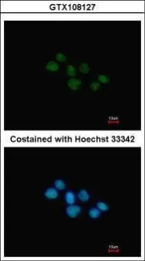
NR1D1 antibody [N1], N-termGTX108127
ApplicationsImmunoFluorescence, ImmunoCytoChemistry
ReactivityHuman
TargetNR1D1
- SizePrice
Product group Antibodies
Anti-NR1D1Y058416
ApplicationsImmunoHistoChemistry
ReactivityHuman, Mouse
- SizePrice
Product group Antibodies
Anti-NR1D1 Antibody101-11130
ApplicationsWestern Blot, ELISA
TargetNR1D1
- SizePrice
Product group Antibodies
NR1D1 Polyclonal AntibodyBS-20222R
ApplicationsFlow Cytometry, ImmunoFluorescence, ImmunoHistoChemistry, ImmunoHistoChemistry Frozen, ImmunoHistoChemistry Paraffin
ReactivityHuman, Mouse, Rat
TargetNR1D1
- SizePrice