


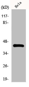
Figure 1. Western blot analysis of NR2F6 using anti-NR2F6 antibody (PB10081). Electrophoresis was performed on a 5-20% SDS-PAGE gel at 70V (Stacking gel) / 90V (Resolving gel) for 2-3 hours. The sample well of each lane was loaded with 30 ug of sample under reducing conditions. Lane 1: rat liver tissue lysates, Lane 2: HELA whole cell lysates. After electrophoresis, proteins were transferred to a nitrocellulose membrane at 150 mA for 50-90 minutes. Blocked the membrane with 5% non-fat milk/TBS for 1.5 hour at RT. The membrane was incubated with rabbit anti-NR2F6 antigen affinity purified polyclonal antibody (Catalog # PB10081) at 0.5 microg/mL overnight at 4°C, then washed with TBS-0.1%Tween 3 times with 5 minutes each and probed with a goat anti-rabbit IgG-HRP secondary antibody at a dilution of 1:5000 for 1.5 hour at RT. The signal is developed using an Enhanced Chemiluminescent detection (ECL) kit (Catalog # EK1002) with Tanon 5200 system. A specific band was detected for NR2F6 at approximately 43 kDa. The expected band size for NR2F6 is at 43 kDa.
Figure 1. Western blot analysis of NR2F6 using anti-NR2F6 antibody (PB10081). Electrophoresis was performed on a 5-20% SDS-PAGE gel at 70V (Stacking gel) / 90V (Resolving gel) for 2-3 hours. The sample well of each lane was loaded with 30 ug of sample under reducing conditions. Lane 1: rat liver tissue lysates, Lane 2: HELA whole cell lysates. After electrophoresis, proteins were transferred to a nitrocellulose membrane at 150 mA for 50-90 minutes. Blocked the membrane with 5% non-fat milk/TBS for 1.5 hour at RT. The membrane was incubated with rabbit anti-NR2F6 antigen affinity purified polyclonal antibody (Catalog # PB10081) at 0.5 microg/mL overnight at 4°C, then washed with TBS-0.1%Tween 3 times with 5 minutes each and probed with a goat anti-rabbit IgG-HRP secondary antibody at a dilution of 1:5000 for 1.5 hour at RT. The signal is developed using an Enhanced Chemiluminescent detection (ECL) kit (Catalog # EK1002) with Tanon 5200 system. A specific band was detected for NR2F6 at approximately 43 kDa. The expected band size for NR2F6 is at 43 kDa.
Anti-NR2F6 Antibody Picoband(r)
PB10081-CARRIER-FREE
ApplicationsWestern Blot
Product group Antibodies
ReactivityBovine, Equine, Human, Monkey, Rat
TargetNR2F6
Overview
- SupplierBoster Bio
- Product NameAnti-NR2F6 Antibody Picoband(r)
- Delivery Days Customer9
- Application Supplier NoteTested Species: In-house tested species with positive results. Other applications have not been tested. Optimal dilutions should be determined by end users.
- ApplicationsWestern Blot
- CertificationResearch Use Only
- ClonalityPolyclonal
- Concentration500 ug/ml
- Gene ID2063
- Target nameNR2F6
- Target descriptionnuclear receptor subfamily 2 group F member 6
- Target synonymsEAR-2, EAR2, ERBAL2, nuclear receptor subfamily 2 group F member 6, ERBA-related gene-2, V-erbA-related protein 2, nuclear receptor V-erbA-related, v-erb-a avian erythroblastic leukemia viral oncogene homolog-like 2
- HostRabbit
- IsotypeIgG
- Protein IDP10588
- Protein NameNuclear receptor subfamily 2 group F member 6
- Scientific DescriptionBoster Bio Anti-NR2F6 Antibody Picoband® catalog # PB10081. Tested in WB applications. This antibody reacts with Human, Rat. The brand Picoband indicates this is a premium antibody that guarantees superior quality, high affinity, and strong signals with minimal background in Western blot applications. Only our best-performing antibodies are designated as Picoband, ensuring unmatched performance.
- ReactivityBovine, Equine, Human, Monkey, Rat
- Storage Instruction-20°C,2°C to 8°C
- UNSPSC12352203
Related products
Product group Antibodies

Anti-NR2F6 [RAB-T39]Ab01822-1.1
ApplicationsFlow Cytometry, ImmunoFluorescence
ReactivityHuman
TargetNR2F6
- SizePrice
Product group Antibodies
Anti-NR2F6 AntibodyA35324
ApplicationsImmunoFluorescence, Western Blot, ImmunoHistoChemistry
ReactivityHuman, Mouse, Rat
- SizePrice
Product group Antibodies

EAR2 / NR2F6 AntibodyLS-C750240
ApplicationsWestern Blot
ReactivityHuman, Mouse, Rat
TargetNR2F6
- SizePrice
Product group Antibodies
NR2F6 AntibodyCSB-PA002242
ApplicationsImmunoFluorescence, Western Blot, ELISA, ImmunoHistoChemistry
ReactivityHuman, Mouse, Rat
TargetNR2F6
- SizePrice
Product group Antibodies
NR2F6 antibodyGTX100255
ApplicationsWestern Blot, ChIP Chromatin ImmunoPrecipitation, ImmunoHistoChemistry, ImmunoHistoChemistry Paraffin
ReactivityHuman, Mouse
TargetNR2F6
- SizePrice