


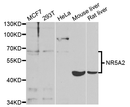
Figure 1. Western blot analysis of NR5A2 using anti-NR5A2 antibody (A01332-1). Electrophoresis was performed on a 5-20% SDS-PAGE gel at 70V (Stacking gel) / 90V (Resolving gel) for 2-3 hours. The sample well of each lane was loaded with 30 ug of sample under reducing conditions. Lane 1: human HepG2 whole cell lysates, Lane 2: human A549 whole cell lysates, Lane 3: human Caco-2 whole cell lysates, Lane 4: human 293T whole cell lysates, Lane 5: human MCF-7 whole cell lysates, Lane 6: human K562 whole cell lysates. After electrophoresis, proteins were transferred to a nitrocellulose membrane at 150 mA for 50-90 minutes. Blocked the membrane with 5% non-fat milk/TBS for 1.5 hour at RT. The membrane was incubated with rabbit anti-NR5A2 antigen affinity purified polyclonal antibody (Catalog # A01332-1) at 0.5 microg/mL overnight at 4°C, then washed with TBS-0.1%Tween 3 times with 5 minutes each and probed with a goat anti-rabbit IgG-HRP secondary antibody at a dilution of 1:5000 for 1.5 hour at RT. The signal is developed using an Enhanced Chemiluminescent detection (ECL) kit (Catalog # EK1002) with Tanon 5200 system. A specific band was detected for NR5A2 at approximately 70 kDa. The expected band size for NR5A2 is at 61 kDa.
. NR5A2 was detected in a paraffin-embedded section of human parotid acinar cell carcinoma tissue. Heat mediated antigen retrieval was performed in EDTA buffer (pH 8.0, epitope retrieval solution). The tissue section was blocked with 10% goat serum. The tissue section was then incubated with 2 microg/ml rabbit anti-NR5A2 Antibody (A01332-1) overnight at 4°C. Peroxidase Conjugated Goat Anti-rabbit IgG was used as secondary antibody and incubated for 30 minutes at 37°C. The tissue section was developed using HRP Conjugated Rabbit IgG Super Vision Assay Kit (Catalog # SV0002) with DAB as the chromogen.")
. NR5A2 was detected in a paraffin-embedded section of human bladder urothelial carcinoma tissue. Heat mediated antigen retrieval was performed in EDTA buffer (pH 8.0, epitope retrieval solution). The tissue section was blocked with 10% goat serum. The tissue section was then incubated with 2 microg/ml rabbit anti-NR5A2 Antibody (A01332-1) overnight at 4°C. Peroxidase Conjugated Goat Anti-rabbit IgG was used as secondary antibody and incubated for 30 minutes at 37°C. The tissue section was developed using HRP Conjugated Rabbit IgG Super Vision Assay Kit (Catalog # SV0002) with DAB as the chromogen.")
. NR5A2 was detected in a paraffin-embedded section of human colorectal adenocarcinoma tissue. Heat mediated antigen retrieval was performed in EDTA buffer (pH 8.0, epitope retrieval solution). The tissue section was blocked with 10% goat serum. The tissue section was then incubated with 2 microg/ml rabbit anti-NR5A2 Antibody (A01332-1) overnight at 4°C. Peroxidase Conjugated Goat Anti-rabbit IgG was used as secondary antibody and incubated for 30 minutes at 37°C. The tissue section was developed using HRP Conjugated Rabbit IgG Super Vision Assay Kit (Catalog # SV0002) with DAB as the chromogen.")
. Overlay histogram showing CACO-2 cells stained with A01332-1 (Blue line). To facilitate intracellular staining, cells were fixed with 4% paraformaldehyde and permeabilized with permeabilization buffer. The cells were blocked with 10% normal goat serum. And then incubated with rabbit anti-NR5A2 Antibody (A01332-1, 1 microg/1x106 cells) for 30 min at 20°C. DyLight®488 conjugated goat anti-rabbit IgG (BA1127, 5-10 microg/1x106 cells) was used as secondary antibody for 30 minutes at 20°C. Isotype control antibody (Green line) was rabbit IgG (1 microg/1x106) used under the same conditions. Unlabelled sample (Red line) was also used as a control.")
Figure 1. Western blot analysis of NR5A2 using anti-NR5A2 antibody (A01332-1). Electrophoresis was performed on a 5-20% SDS-PAGE gel at 70V (Stacking gel) / 90V (Resolving gel) for 2-3 hours. The sample well of each lane was loaded with 30 ug of sample under reducing conditions. Lane 1: human HepG2 whole cell lysates, Lane 2: human A549 whole cell lysates, Lane 3: human Caco-2 whole cell lysates, Lane 4: human 293T whole cell lysates, Lane 5: human MCF-7 whole cell lysates, Lane 6: human K562 whole cell lysates. After electrophoresis, proteins were transferred to a nitrocellulose membrane at 150 mA for 50-90 minutes. Blocked the membrane with 5% non-fat milk/TBS for 1.5 hour at RT. The membrane was incubated with rabbit anti-NR5A2 antigen affinity purified polyclonal antibody (Catalog # A01332-1) at 0.5 microg/mL overnight at 4°C, then washed with TBS-0.1%Tween 3 times with 5 minutes each and probed with a goat anti-rabbit IgG-HRP secondary antibody at a dilution of 1:5000 for 1.5 hour at RT. The signal is developed using an Enhanced Chemiluminescent detection (ECL) kit (Catalog # EK1002) with Tanon 5200 system. A specific band was detected for NR5A2 at approximately 70 kDa. The expected band size for NR5A2 is at 61 kDa.
Anti-NR5A2 Antibody Picoband(r)
A01332-1-CARRIER-FREE
ApplicationsFlow Cytometry, Western Blot, ELISA, ImmunoHistoChemistry
Product group Antibodies
ReactivityHuman
TargetNR5A2
Overview
- SupplierBoster Bio
- Product NameAnti-NR5A2 Antibody Picoband(r)
- Delivery Days Customer9
- ApplicationsFlow Cytometry, Western Blot, ELISA, ImmunoHistoChemistry
- CertificationResearch Use Only
- ClonalityPolyclonal
- Concentration500 ug/ml
- Gene ID2494
- Target nameNR5A2
- Target descriptionnuclear receptor subfamily 5 group A member 2
- Target synonymsB1F, B1F2, CPF, FTF, FTZ-F1, FTZ-F1beta, LRH-1, LRH1, hB1F-2, nuclear receptor subfamily 5 group A member 2, CYP7A promoter-binding factor, b1-binding factor, hepatocyte transcription factor which activates enhancer II of hepatitis B virus, fetoprotein-alpha 1 (AFP) transcription factor, hepatocytic transcription factor hB1F-3
- HostRabbit
- IsotypeIgG
- Protein IDO00482
- Protein NameNuclear receptor subfamily 5 group A member 2
- Scientific DescriptionBoster Bio Anti-NR5A2 Antibody Picoband® catalog # A01332-1. Tested in ELISA, IHC, WB, Flow Cytometry applications. This antibody reacts with Human. The brand Picoband indicates this is a premium antibody that guarantees superior quality, high affinity, and strong signals with minimal background in Western blot applications. Only our best-performing antibodies are designated as Picoband, ensuring unmatched performance.
- ReactivityHuman
- Storage Instruction-20°C,2°C to 8°C
- UNSPSC12352203
Related products
Product group Antibodies

Anti-NR5A2 [RAB-T41]Ab01824-1.1
ApplicationsFlow Cytometry, ImmunoFluorescence
ReactivityHuman
TargetNR5A2
- SizePrice
Product group Antibodies
Anti-NR5A2 Antibody144-62861
ApplicationsWestern Blot
ReactivityHuman, Mouse
TargetNR5A2
- SizePrice
Product group Antibodies
References
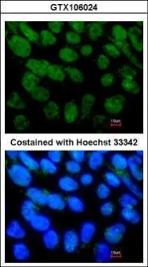
NR5A2 antibody [N2C3]GTX106024
ApplicationsImmunoFluorescence, Western Blot, ImmunoCytoChemistry
ReactivityHuman
TargetNR5A2
- SizePrice
Product group Antibodies
NR5A2 Polyclonal AntibodyCAC14743
ApplicationsWestern Blot, ELISA, ImmunoHistoChemistry
ReactivityMouse
TargetNR5A2
- SizePrice
Product group Antibodies
NR5A2 Recombinant Antibody, AbBy Fluor-488 ConjugatedBSM-61508R-BF488
ApplicationsWestern Blot
ReactivityHuman, Mouse, Rat
TargetNR5A2
- SizePrice
Product group Antibodies
Anti-NR5A2 AntibodyA35305
ApplicationsImmunoFluorescence, Western Blot, ImmunoHistoChemistry
ReactivityHuman, Mouse, Rat
- SizePrice
Product group Antibodies
NR5A2 AntibodyCSB-PA016066LA01HU
ApplicationsWestern Blot, ELISA, ImmunoHistoChemistry
ReactivityHuman, Mouse
TargetNR5A2
- SizePrice
Product group Antibodies

Goat anti-NR5A2 / LRH1EB12283
ApplicationsWestern Blot, ELISA
ReactivityBovine, Canine, Human, Mouse, Porcine, Rat
TargetNR5A2
- SizePrice