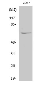
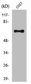
Bio-Connect
Anti-NUB1 Antibody

Anti-NUB1 Antibody

A36031
Antibodies.com
ApplicationsWestern Blot, ELISA, ImmunoHistoChemistry
Product group Antibodies
Sign in to order and to see your custom pricing.
Large volume orders?
Order with a bulk request

Overview

  • Supplier
    Antibodies.com
  • Product Name
    Anti-NUB1 Antibody
  • Delivery Days Customer
    7
  • Applications
    Western Blot, ELISA, ImmunoHistoChemistry
  • Certification
    Research Use Only
  • Clonality
    Polyclonal
  • Concentration
    1 mg/ml
  • Conjugate
    Unconjugated
  • Host
    Rabbit
  • Scientific Description
    Rabbit polyclonal antibody to NUB1
  • UNSPSC
    12352203

Related products

Immunofluorescent staining of human cell line MCF7 shows localization to nucleus & nucleoli.
Product group Antibodies
Atlas Antibodies
ApplicationsImmunoCytoChemistry
TargetNUB1
  • SizePrice
Product group Antibodies
LifeSpan BioSciences
ApplicationsWestern Blot, ELISA, ImmunoHistoChemistry
TargetNUB1
  • SizePrice
Product group Antibodies
RayBiotech
ApplicationsWestern Blot, ImmunoHistoChemistry, ImmunoHistoChemistry Paraffin
TargetNUB1
  • SizePrice
Product group Antibodies
GeneTex
ApplicationsImmunoFluorescence, Western Blot, ImmunoCytoChemistry
TargetNUB1
  • SizePrice
Figure 1. Western blot analysis of NUB1 using anti-NUB1 antibody (A06089-2). Electrophoresis was performed on a 5-20% SDS-PAGE gel at 70V (Stacking gel) / 90V (Resolving gel) for 2-3 hours. The sample well of each lane was loaded with 30 ug of sample under reducing conditions. Lane 1: human Hela whole cell lysates, Lane 2: human Colo320 whole cell lysates, Lane 3: human HepG2 whole cell lysates, Lane 4: human K562 whole cell lysates, Lane 5: human Raji whole cell lysates, Lane 6: monkey COS-7 whole cell lysates, Lane 8: human MCF-7 whole cell lysates, Lane 9: human A549 whole cell lysates, Lane 10: rat liver tissue lysates, Lane 11: rat PC-12 whole cell lysates, Lane 12: mouse NIH/3T3 whole cell lysates. After electrophoresis, proteins were transferred to a nitrocellulose membrane at 150 mA for 50-90 minutes. Blocked the membrane with 5% non-fat milk/TBS for 1.5 hour at RT. The membrane was incubated with rabbit anti-NUB1 antigen affinity purified polyclonal antibody (Catalog # A06089-2) at 0.25 microg/mL overnight at 4°C, then washed with TBS-0.1%Tween 3 times with 5 minutes each and probed with a goat anti-rabbit IgG-HRP secondary antibody at a dilution of 1:5000 for 1.5 hour at RT. The signal is developed using an Enhanced Chemiluminescent detection (ECL) kit (Catalog # EK1002) with Tanon 5200 system. A specific band was detected for NUB1 at approximately 75 kDa. The expected band size for NUB1 is at 71 kDa.
Product group Antibodies
Boster Bio
Anti-NUB1 Antibody Picoband(r)A06089-2-CARRIER-FREE
ApplicationsFlow Cytometry, ImmunoFluorescence, Western Blot, ELISA, ImmunoCytoChemistry, ImmunoHistoChemistry
TargetNUB1
  • SizePrice
Western Blot analysis of COS7 cells using NUB1 Polyclonal Antibody
Product group Antibodies
Cusabio
NUB1 AntibodyCSB-PA003491
ApplicationsWestern Blot, ELISA, ImmunoHistoChemistry
ReactivityHuman, Monkey, Mouse
TargetNUB1
  • SizePrice