


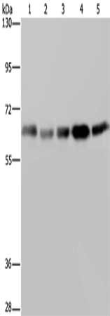
Figure 1. Western blot analysis of Nucleostemin/GNL3 using anti-Nucleostemin/GNL3 antibody (A03283-3). Electrophoresis was performed on a 5-20% SDS-PAGE gel at 70V (Stacking gel) / 90V (Resolving gel) for 2-3 hours. The sample well of each lane was loaded with 30 ug of sample under reducing conditions. Lane 1: human Hela whole cell lysates, Lane 2: human K562 whole cell lysates, Lane 3: human PC-3 whole cell lysates, Lane 4: human Jurkat whole cell lysates, Lane 5: rat C6 whole cell lysates, Lane 6: mouse NIH/3T3 whole cell lysates. After electrophoresis, proteins were transferred to a nitrocellulose membrane at 150 mA for 50-90 minutes. Blocked the membrane with 5% non-fat milk/TBS for 1.5 hour at RT. The membrane was incubated with rabbit anti-Nucleostemin/GNL3 antigen affinity purified polyclonal antibody (Catalog # A03283-3) at 0.5 microg/mL overnight at 4°C, then washed with TBS-0.1%Tween 3 times with 5 minutes each and probed with a goat anti-rabbit IgG-HRP secondary antibody at a dilution of 1:5000 for 1.5 hour at RT. The signal is developed using an Enhanced Chemiluminescent detection (ECL) kit (Catalog # EK1002) with Tanon 5200 system. A specific band was detected for Nucleostemin/GNL3 at approximately 65 kDa. The expected band size for Nucleostemin/GNL3 is at 65 kDa.
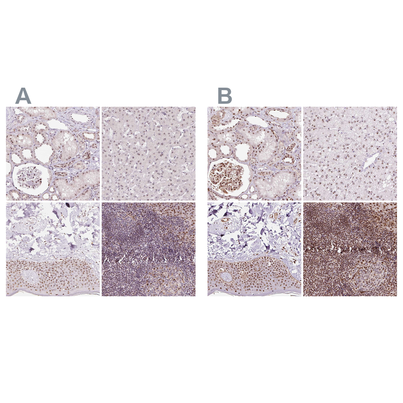
. Nucleostemin/GNL3 was detected in a paraffin-embedded section of human appendiceal adenocarcinoma tissue. Heat mediated antigen retrieval was performed in EDTA buffer (pH 8.0, epitope retrieval solution). The tissue section was blocked with 10% goat serum. The tissue section was then incubated with 2 microg/ml rabbit anti-Nucleostemin/GNL3 Antibody (A03283-3) overnight at 4°C. Biotinylated goat anti-rabbit IgG was used as secondary antibody and incubated for 30 minutes at 37°C. The tissue section was developed using Strepavidin-Biotin-Complex (SABC) (Catalog # SA1022) with DAB as the chromogen.")
. Nucleostemin/GNL3 was detected in a paraffin-embedded section of human breast cancer tissue. Heat mediated antigen retrieval was performed in EDTA buffer (pH 8.0, epitope retrieval solution). The tissue section was blocked with 10% goat serum. The tissue section was then incubated with 2 microg/ml rabbit anti-Nucleostemin/GNL3 Antibody (A03283-3) overnight at 4°C. Biotinylated goat anti-rabbit IgG was used as secondary antibody and incubated for 30 minutes at 37°C. The tissue section was developed using Strepavidin-Biotin-Complex (SABC) (Catalog # SA1022) with DAB as the chromogen.")
. Nucleostemin/GNL3 was detected in a paraffin-embedded section of human colonic adenocarcinoma tissue. Heat mediated antigen retrieval was performed in EDTA buffer (pH 8.0, epitope retrieval solution). The tissue section was blocked with 10% goat serum. The tissue section was then incubated with 2 microg/ml rabbit anti-Nucleostemin/GNL3 Antibody (A03283-3) overnight at 4°C. Biotinylated goat anti-rabbit IgG was used as secondary antibody and incubated for 30 minutes at 37°C. The tissue section was developed using Strepavidin-Biotin-Complex (SABC) (Catalog # SA1022) with DAB as the chromogen.")
. Nucleostemin/GNL3 was detected in a paraffin-embedded section of human gall bladder adenosquamous carcinoma tissue. Heat mediated antigen retrieval was performed in EDTA buffer (pH 8.0, epitope retrieval solution). The tissue section was blocked with 10% goat serum. The tissue section was then incubated with 2 microg/ml rabbit anti-Nucleostemin/GNL3 Antibody (A03283-3) overnight at 4°C. Biotinylated goat anti-rabbit IgG was used as secondary antibody and incubated for 30 minutes at 37°C. The tissue section was developed using Strepavidin-Biotin-Complex (SABC) (Catalog # SA1022) with DAB as the chromogen.")
. Nucleostemin/GNL3 was detected in an immunocytochemical section of HEP3B cells. Enzyme antigen retrieval was performed using IHC enzyme antigen retrieval reagent (AR0022) for 15 mins. The cells were blocked with 10% goat serum. And then incubated with 5 microg/mL rabbit anti-Nucleostemin/GNL3 Antibody (A03283-3) overnight at 4°C. DyLight®488 Conjugated Goat Anti-Rabbit IgG (BA1127) was used as secondary antibody at 1:100 dilution and incubated for 30 minutes at 37°C. The tissue section was developed using Phalloidin-iFluor 555 Conjugated. Visualize using a fluorescence microscope and filter sets appropriate for the label used.")
. Overlay histogram showing Hela cells stained with A03283-3 (Blue line). To facilitate intracellular staining, cells were fixed with 4% paraformaldehyde and permeabilized with permeabilization buffer. The cells were blocked with 10% normal goat serum. And then incubated with rabbit anti-Nucleostemin/GNL3 Antibody (A03283-3, 1 microg/1x106 cells) for 30 min at 20°C. DyLight®488 conjugated goat anti-rabbit IgG (BA1127, 5-10 microg/1x106 cells) was used as secondary antibody for 30 minutes at 20°C. Isotype control antibody (Green line) was rabbit IgG (1 microg/1x106) used under the same conditions. Unlabelled sample without incubation with primary antibody and secondary antibody (Red line) was used as a blank control.")
Figure 1. Western blot analysis of Nucleostemin/GNL3 using anti-Nucleostemin/GNL3 antibody (A03283-3). Electrophoresis was performed on a 5-20% SDS-PAGE gel at 70V (Stacking gel) / 90V (Resolving gel) for 2-3 hours. The sample well of each lane was loaded with 30 ug of sample under reducing conditions. Lane 1: human Hela whole cell lysates, Lane 2: human K562 whole cell lysates, Lane 3: human PC-3 whole cell lysates, Lane 4: human Jurkat whole cell lysates, Lane 5: rat C6 whole cell lysates, Lane 6: mouse NIH/3T3 whole cell lysates. After electrophoresis, proteins were transferred to a nitrocellulose membrane at 150 mA for 50-90 minutes. Blocked the membrane with 5% non-fat milk/TBS for 1.5 hour at RT. The membrane was incubated with rabbit anti-Nucleostemin/GNL3 antigen affinity purified polyclonal antibody (Catalog # A03283-3) at 0.5 microg/mL overnight at 4°C, then washed with TBS-0.1%Tween 3 times with 5 minutes each and probed with a goat anti-rabbit IgG-HRP secondary antibody at a dilution of 1:5000 for 1.5 hour at RT. The signal is developed using an Enhanced Chemiluminescent detection (ECL) kit (Catalog # EK1002) with Tanon 5200 system. A specific band was detected for Nucleostemin/GNL3 at approximately 65 kDa. The expected band size for Nucleostemin/GNL3 is at 65 kDa.
Anti-Nucleostemin/GNL3 Antibody Picoband(r)
A03283-3-CARRIER-FREE
ApplicationsFlow Cytometry, ImmunoFluorescence, Western Blot, ELISA, ImmunoCytoChemistry, ImmunoHistoChemistry
Product group Antibodies
ReactivityHuman, Mouse, Rat
TargetGNL3
Overview
- SupplierBoster Bio
- Product NameAnti-Nucleostemin/GNL3 Antibody Picoband(r)
- Delivery Days Customer9
- ApplicationsFlow Cytometry, ImmunoFluorescence, Western Blot, ELISA, ImmunoCytoChemistry, ImmunoHistoChemistry
- CertificationResearch Use Only
- ClonalityPolyclonal
- Concentration500 ug/ml
- Gene ID26354
- Target nameGNL3
- Target descriptionG protein nucleolar 3
- Target synonymsC77032, E2IG3, NNP47, NS, Nug1, guanine nucleotide-binding protein-like 3, E2-induced gene 3 protein, estradiol-induced nucleotide binding protein, guanine nucleotide binding protein-like 3 (nucleolar), novel nucleolar protein 47, nucleolar GTP-binding protein 3, nucleostemin
- HostRabbit
- IsotypeIgG
- Protein IDQ9BVP2
- Protein NameGuanine nucleotide-binding protein-like 3
- Scientific DescriptionBoster Bio Anti-Nucleostemin/GNL3 Antibody Picoband® catalog # A03283-3. Tested in ELISA, Flow Cytometry, IF, IHC, ICC, WB applications. This antibody reacts with Human, Mouse, Rat. The brand Picoband indicates this is a premium antibody that guarantees superior quality, high affinity, and strong signals with minimal background in Western blot applications. Only our best-performing antibodies are designated as Picoband, ensuring unmatched performance.
- ReactivityHuman, Mouse, Rat
- Storage Instruction-20°C,2°C to 8°C
- UNSPSC12352203
Related products
Product group Antibodies
ApplicationsWestern Blot, ELISA, ImmunoHistoChemistry
ReactivityHuman
- SizePrice
Product group Antibodies
Anti-GNL3 Antibody144-06459
ApplicationsImmunoFluorescence, ImmunoPrecipitation, Western Blot
ReactivityHuman
TargetGNL3
- SizePrice
Product group Antibodies
GNL3 Recombinant AntibodyBSM-61946R
ApplicationsFlow Cytometry, ImmunoFluorescence, Western Blot, ImmunoCytoChemistry
ReactivityHuman
TargetGNL3
- SizePrice
Product group Antibodies

Goat anti-GNL3 (aa115-126)EB12563
ApplicationsWestern Blot, ELISA, ImmunoHistoChemistry
ReactivityHuman
TargetGNL3
- SizePrice
Product group Antibodies
GNL3 AntibodyCSB-PA184930
ApplicationsWestern Blot, ELISA
ReactivityHuman
TargetGNL3
- SizePrice
Product group Antibodies

GNL3 / NS / Nucleostemin AntibodyLS-C401900
ApplicationsWestern Blot, ELISA, ImmunoHistoChemistry
ReactivityHuman
TargetGNL3
- SizePrice
Product group Antibodies
Nucleostemin antibody, InternalGTX44808
ApplicationsWestern Blot
ReactivityHuman
TargetGNL3
- SizePrice
Product group Antibodies
Anti-GNL3 AntibodyHPA036742
ApplicationsWestern Blot, ImmunoHistoChemistry
ReactivityHuman
TargetGNL3
- SizePrice
Product group Antibodies
Anti-GNL3 AntibodyCAB6459
ApplicationsImmunoFluorescence, ImmunoPrecipitation, Western Blot, ELISA, ImmunoCytoChemistry
ReactivityHuman
TargetGNL3
- SizePrice