


Immunohistochemical staining of human kidney shows strong cytoplasmic positivity in granular pattern in tubular cells.
Immunohistochemical staining of human kidney shows strong cytoplasmic positivity in granular pattern in tubular cells.
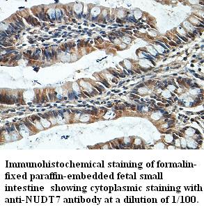
Anti-NUDT7 Antibody
HPA042042
ApplicationsImmunoHistoChemistry
Product group Antibodies
ReactivityHuman
TargetNUDT7
Overview
- SupplierAtlas Antibodies
- Product NameAnti-NUDT7 Antibody
- Delivery Days Customer4
- ApplicationsImmunoHistoChemistry
- CertificationResearch Use Only
- ClonalityPolyclonal
- ConjugateUnconjugated
- Gene ID283927
- Target nameNUDT7
- Target descriptionnudix hydrolase 7
- Target synonymsperoxisomal coenzyme A diphosphatase NUDT7, nudix (nucleoside diphosphate linked moiety X)-type motif 7
- HostRabbit
- IsotypeIgG
- Protein IDP0C024
- Protein NamePeroxisomal coenzyme A diphosphatase NUDT7
- Scientific DescriptionRecombinant Protein Epitope Signature Tag (PrEST) antigen sequence
- ReactivityHuman
- Storage Instruction-20°C,2°C to 8°C
- UNSPSC41116161
Datasheet
MSDS
Related products
Product group Antibodies
Anti-NUDT7 AntibodyA38160
ApplicationsWestern Blot, ImmunoHistoChemistry
ReactivityHuman
- SizePrice
Product group Antibodies

NUDT7 AntibodyLS-C137239
ApplicationsWestern Blot, ImmunoHistoChemistry
ReactivityHuman
TargetNUDT7
- SizePrice
Product group Antibodies
NUDT7 AntibodyCSB-PA890440XA01DOA
ApplicationsWestern Blot, ELISA
ReactivityPlant
TargetNUDT7
- SizePrice
Product group Antibodies
Anti-NUDT7Y058467
ApplicationsWestern Blot, ImmunoHistoChemistry
ReactivityHuman
- SizePrice

Product group Antibodies
Anti-NUDT7 Antibody Picoband(r)A10663-1-CARRIER-FREE
ApplicationsFlow Cytometry, Western Blot, ELISA, ImmunoHistoChemistry
ReactivityHuman, Mouse, Rat
TargetNUDT7
- SizePrice