


Immunofluorescent staining of human cell line U-2 OS shows localization to nucleus.
Immunofluorescent staining of human cell line U-2 OS shows localization to nucleus.
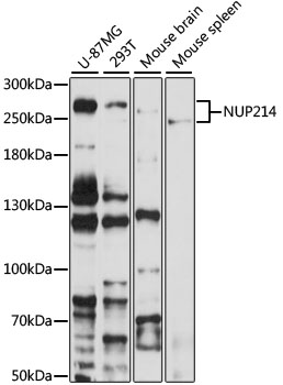
Anti-NUP214 Antibody
HPA048789
ApplicationsImmunoCytoChemistry
Product group Antibodies
ReactivityHuman
TargetNUP214
Overview
- SupplierAtlas Antibodies
- Product NameAnti-NUP214 Antibody
- Delivery Days Customer4
- ApplicationsImmunoCytoChemistry
- CertificationResearch Use Only
- ClonalityPolyclonal
- ConjugateUnconjugated
- Gene ID8021
- Target nameNUP214
- Target descriptionnucleoporin 214
- Target synonymsCAIN, CAN, IIAE9, nuclear pore complex protein Nup214, CAN protein, putative oncogene, nucleoporin 214kDa
- HostRabbit
- IsotypeIgG
- Protein IDP35658
- Protein NameNuclear pore complex protein Nup214
- Scientific DescriptionRecombinant Protein Epitope Signature Tag (PrEST) antigen sequence
- ReactivityHuman
- Storage Instruction-20°C,2°C to 8°C
- UNSPSC41116161
Datasheet
MSDS
Related products
Product group Antibodies
Anti-NUP214 AntibodyA12242
ApplicationsImmunoFluorescence, Western Blot, ImmunoCytoChemistry
ReactivityHuman, Mouse, Rat
- SizePrice

Product group Antibodies
Anti-NUP214 Antibody Picoband(r)A02408-CARRIER-FREE
ApplicationsFlow Cytometry, ImmunoFluorescence, Western Blot, ELISA, ImmunoCytoChemistry
ReactivityHuman, Mouse
TargetNUP214
- SizePrice
Product group Antibodies
Anti-NUP214 Antibody144-66594
ApplicationsImmunoFluorescence, Western Blot
ReactivityHuman, Mouse, Rat
TargetNUP214
- SizePrice
Product group Antibodies
NUP214 AntibodyCSB-PA973556XA01DOA
ApplicationsWestern Blot, ELISA
ReactivityPlant
- SizePrice
Product group Antibodies

NUP214 / CAN AntibodyLS-C409893
ApplicationsWestern Blot
ReactivityHuman, Mouse
TargetNUP214
- SizePrice
Product group Antibodies
Anti-NUP214 AntibodyCAB8357
ApplicationsImmunoFluorescence, Western Blot, ELISA, ImmunoCytoChemistry
ReactivityHuman
TargetNUP214
- SizePrice