


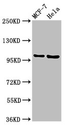
Figure 1. Western blot analysis of NUP98 using anti-NUP98 antibody (A01301-1). Electrophoresis was performed on a 5-20% SDS-PAGE gel at 70V (Stacking gel) / 90V (Resolving gel) for 2-3 hours. The sample well of each lane was loaded with 30 ug of sample under reducing conditions. Lane 1: human Caco-2 whole cell lysates, Lane 2: human Hela whole cell lysates, Lane 3: human K562 whole cell lysates, Lane 4: monkey COS-7 whole cell lysates, Lane 5: rat NRK whole cell lysates, Lane 6: rat PC-12 whole cell lysates, Lane 7: mouse RAW264.7 whole cell lysates, Lane 8: mouse NIH/3T3 whole cell lysates. After electrophoresis, proteins were transferred to a nitrocellulose membrane at 150 mA for 50-90 minutes. Blocked the membrane with 5% non-fat milk/TBS for 1.5 hour at RT. The membrane was incubated with rabbit anti-NUP98 antigen affinity purified polyclonal antibody (Catalog # A01301-1) at 0.5 microg/mL overnight at 4°C, then washed with TBS-0.1%Tween 3 times with 5 minutes each and probed with a goat anti-rabbit IgG-HRP secondary antibody at a dilution of 1:5000 for 1.5 hour at RT. The signal is developed using an Enhanced Chemiluminescent detection (ECL) kit (Catalog # EK1002) with Tanon 5200 system. A specific band was detected for NUP98 at approximately 98,105 kDa. The expected band size for NUP98 is at 198 kDa.
. NUP98 was detected in an immunocytochemical section of A549 cells. Enzyme antigen retrieval was performed using IHC enzyme antigen retrieval reagent (AR0022) for 15 mins. The cells were blocked with 10% goat serum. And then incubated with 5 microg/mL rabbit anti-NUP98 Antibody (A01301-1) overnight at 4°C. Cy3 Conjugated Goat Anti-Rabbit IgG (BA1032) was used as secondary antibody at 1:500 dilution and incubated for 30 minutes at 37°C. The section was counterstained with DAPI. Visualize using a fluorescence microscope and filter sets appropriate for the label used.")
. Overlay histogram showing K562 cells stained with A01301-1 (Blue line). To facilitate intracellular staining, cells were fixed with 4% paraformaldehyde and permeabilized with permeabilization buffer. The cells were blocked with 10% normal goat serum. And then incubated with rabbit anti-NUP98 Antibody (A01301-1, 1 microg/1x106 cells) for 30 min at 20°C. DyLight®488 conjugated goat anti-rabbit IgG (BA1127, 5-10 microg/1x106 cells) was used as secondary antibody for 30 minutes at 20°C. Isotype control antibody (Green line) was rabbit IgG (1 microg/1x106) used under the same conditions. Unlabelled sample (Red line) was also used as a control.")
Figure 1. Western blot analysis of NUP98 using anti-NUP98 antibody (A01301-1). Electrophoresis was performed on a 5-20% SDS-PAGE gel at 70V (Stacking gel) / 90V (Resolving gel) for 2-3 hours. The sample well of each lane was loaded with 30 ug of sample under reducing conditions. Lane 1: human Caco-2 whole cell lysates, Lane 2: human Hela whole cell lysates, Lane 3: human K562 whole cell lysates, Lane 4: monkey COS-7 whole cell lysates, Lane 5: rat NRK whole cell lysates, Lane 6: rat PC-12 whole cell lysates, Lane 7: mouse RAW264.7 whole cell lysates, Lane 8: mouse NIH/3T3 whole cell lysates. After electrophoresis, proteins were transferred to a nitrocellulose membrane at 150 mA for 50-90 minutes. Blocked the membrane with 5% non-fat milk/TBS for 1.5 hour at RT. The membrane was incubated with rabbit anti-NUP98 antigen affinity purified polyclonal antibody (Catalog # A01301-1) at 0.5 microg/mL overnight at 4°C, then washed with TBS-0.1%Tween 3 times with 5 minutes each and probed with a goat anti-rabbit IgG-HRP secondary antibody at a dilution of 1:5000 for 1.5 hour at RT. The signal is developed using an Enhanced Chemiluminescent detection (ECL) kit (Catalog # EK1002) with Tanon 5200 system. A specific band was detected for NUP98 at approximately 98,105 kDa. The expected band size for NUP98 is at 198 kDa.
Anti-NUP98 Antibody Picoband(r)
A01301-1-CARRIER-FREE
ApplicationsFlow Cytometry, ImmunoFluorescence, Western Blot, ELISA, ImmunoCytoChemistry
Product group Antibodies
ReactivityHuman, Monkey, Mouse, Rat
TargetNUP98
Overview
- SupplierBoster Bio
- Product NameAnti-NUP98 Antibody Picoband(r)
- Delivery Days Customer9
- ApplicationsFlow Cytometry, ImmunoFluorescence, Western Blot, ELISA, ImmunoCytoChemistry
- CertificationResearch Use Only
- ClonalityPolyclonal
- Concentration500 ug/ml
- Gene ID4928
- Target nameNUP98
- Target descriptionnucleoporin 98 and 96 precursor
- Target synonymsADIR2, NUP196, NUP96, Nup98-96, nuclear pore complex protein Nup98-Nup96, nuclear pore complex protein Nup98, GLFG-repeat containing nucleoporin, NUP98/PHF23 fusion 2 protein, Nup98-Nup96, nucleoporin 96, nucleoporin 98kD, nucleoporin 98kDa
- HostRabbit
- IsotypeIgG
- Protein IDP52948
- Protein NameNuclear pore complex protein Nup98-Nup96
- Scientific DescriptionBoster Bio Anti-NUP98 Antibody Picoband® catalog # A01301-1. Tested in WB, ICC/IF, Flow Cytometry, ELISA applications. This antibody reacts with Human, Monkey, Mouse, Rat. The brand Picoband indicates this is a premium antibody that guarantees superior quality, high affinity, and strong signals with minimal background in Western blot applications. Only our best-performing antibodies are designated as Picoband, ensuring unmatched performance.
- ReactivityHuman, Monkey, Mouse, Rat
- Storage Instruction-20°C,2°C to 8°C
- UNSPSC12352203
Related products
Product group Antibodies
NUP98 AntibodyCSB-PA016209LA01HU
ApplicationsImmunoFluorescence, ImmunoPrecipitation, Western Blot, ELISA
ReactivityHuman
TargetNUP98
- SizePrice
Product group Antibodies
Anti-NUP98 AntibodyA32479
ApplicationsImmunoPrecipitation, Western Blot
ReactivityHuman
- SizePrice
Product group Antibodies

NUP98 AntibodyLS-C768691
ApplicationsWestern Blot, ELISA
ReactivityHuman, Mouse, Rat
TargetNUP98
- SizePrice

Product group Antibodies
Anti-NUP98 AntibodyHPA074810
ApplicationsImmunoCytoChemistry
ReactivityHuman
TargetNUP98
- SizePrice
Product group Antibodies
NUP98 Polyclonal AntibodyCAC13141
ApplicationsImmunoFluorescence, ImmunoPrecipitation, Western Blot, ELISA
TargetNUP98
- SizePrice
Product group Antibodies
NUP98 Recombinant AntibodyBSM-61197R
ApplicationsImmunoFluorescence, Western Blot, ImmunoCytoChemistry, ImmunoHistoChemistry, ImmunoHistoChemistry Frozen, ImmunoHistoChemistry Paraffin
TargetNUP98
- SizePrice
Product group Antibodies
NUP98 antibody [N1N2], N-termGTX102699
ApplicationsWestern Blot
ReactivityHuman, Monkey
TargetNUP98
- SizePrice
Product group Antibodies
Anti-NUP98 Antibody144-00530
ApplicationsWestern Blot
ReactivityHuman, Mouse
TargetNUP98
- SizePrice