


Immunohistochemical staining of human esophagus shows moderate nuclear and nucleolar positivity in squamous epithelial cells.
![Lane 1: Marker [kDa] 230, 130, 95, 72, 56, 36, 28, 17, 11. Lane 2: Human cell line RT-4. Lane 3: Human cell line U-251MG sp](https://atlasantibodies.s3.amazonaws.com/images/wb/hpa028207-wb-1.jpg "Lane 1: Marker [kDa] 230, 130, 95, 72, 56, 36, 28, 17, 11. Lane 2: Human cell line RT-4. Lane 3: Human cell line U-251MG sp")
Immunohistochemical staining of human esophagus shows moderate nuclear and nucleolar positivity in squamous epithelial cells.
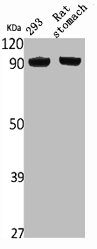
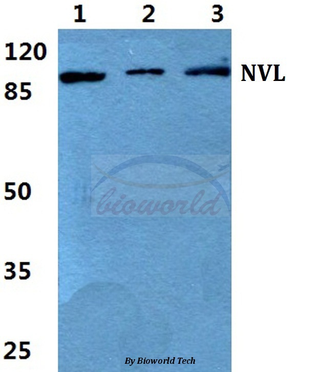
Anti-NVL Antibody
HPA028207
ApplicationsWestern Blot, ImmunoHistoChemistry
Product group Antibodies
ReactivityHuman, Mouse, Rat
TargetNVL
Overview
- SupplierAtlas Antibodies
- Product NameAnti-NVL Antibody
- Delivery Days Customer4
- ApplicationsWestern Blot, ImmunoHistoChemistry
- CertificationResearch Use Only
- ClonalityPolyclonal
- ConjugateUnconjugated
- Gene ID4931
- Target nameNVL
- Target descriptionnuclear VCP like
- Target synonymsNVL2, nuclear valosin-containing protein-like, NVLp
- HostRabbit
- IsotypeIgG
- Protein IDO15381
- Protein NameNuclear valosin-containing protein-like
- Scientific DescriptionRecombinant Protein Epitope Signature Tag (PrEST) antigen sequence
- ReactivityHuman, Mouse, Rat
- Storage Instruction-20°C,2°C to 8°C
- UNSPSC41116161
Datasheet
MSDS
Related products
Product group Antibodies
NVL AntibodyCSB-PA004660
ApplicationsWestern Blot, ELISA
ReactivityHuman, Mouse, Rat
TargetNVL
- SizePrice

Product group Antibodies
Anti-NVL Antibody Picoband(r)A05446-3-CARRIER-FREE
ApplicationsFlow Cytometry, ImmunoFluorescence, Western Blot, ELISA, ImmunoCytoChemistry, ImmunoHistoChemistry
ReactivityHuman, Rat
TargetNVL
- SizePrice
Product group Antibodies
Anti-NVL AntibodyA28073
ApplicationsWestern Blot
ReactivityHuman, Mouse, Rat
- SizePrice
Product group Antibodies

NVL AntibodyLS-C830839
ApplicationsWestern Blot, ELISA, ImmunoHistoChemistry
ReactivityHuman, Mouse
TargetNVL
- SizePrice

Product group Antibodies
Anti-NVL AntibodyHPA028219
ApplicationsImmunoCytoChemistry
ReactivityHuman
TargetNVL
- SizePrice

Product group Antibodies
Anti-NVL-25ulHPA028224
ApplicationsImmunoCytoChemistry, ImmunoHistoChemistry
ReactivityHuman
- SizePrice