

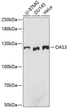


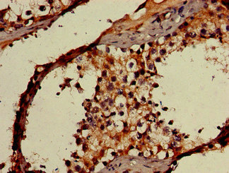
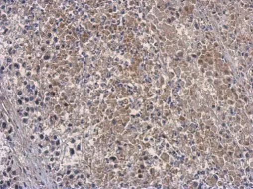
Anti-OAS3 Antibody
A10720
ApplicationsImmunoFluorescence, Western Blot, ImmunoCytoChemistry, ImmunoHistoChemistry
Product group Antibodies
ReactivityHuman, Mouse, Rat
Overview
- SupplierAntibodies.com
- Product NameAnti-OAS3 Antibody
- Delivery Days Customer7
- ApplicationsImmunoFluorescence, Western Blot, ImmunoCytoChemistry, ImmunoHistoChemistry
- CertificationResearch Use Only
- ClonalityPolyclonal
- ConjugateUnconjugated
- HostRabbit
- IsotypeIgG
- Scientific DescriptionRabbit polyclonal antibody to OAS3.
- ReactivityHuman, Mouse, Rat
- UNSPSC12352203
Related products

Product group Antibodies
Anti-OAS3 Antibody Picoband(r)A05032-1-CARRIER-FREE
ApplicationsFlow Cytometry, ImmunoFluorescence, Western Blot, ELISA, ImmunoCytoChemistry
ReactivityHuman
TargetOAS3
- SizePrice
Product group Antibodies
Anti-OAS3 Antibody144-61477
ApplicationsWestern Blot
ReactivityHuman
TargetOAS3
- SizePrice
Product group Antibodies
OAS3 Polyclonal AntibodyBS-7344R
ApplicationsImmunoFluorescence, Western Blot, ELISA, ImmunoCytoChemistry, ImmunoHistoChemistry, ImmunoHistoChemistry Frozen, ImmunoHistoChemistry Paraffin
ReactivityHuman, Mouse, Rat
TargetOAS3
- SizePrice

Product group Antibodies
Anti-OAS3 AntibodyHPA041253
ApplicationsWestern Blot, ImmunoCytoChemistry, ImmunoHistoChemistry
ReactivityHuman
TargetOAS3
- SizePrice
Product group Antibodies
OAS3 AntibodyCSB-PA897591LA01HU
ApplicationsImmunoFluorescence, ELISA, ImmunoHistoChemistry
ReactivityHuman
TargetOAS3
- SizePrice
Product group Antibodies

Goat anti-OAS3 AntibodyEB12375
ApplicationsWestern Blot, ELISA
ReactivityHuman
TargetOAS3
- SizePrice
Product group Antibodies

OAS3 / p100 AntibodyLS-C411016
ApplicationsWestern Blot
ReactivityHuman
TargetOAS3
- SizePrice
Product group Antibodies
OAS3 antibodyGTX118059
ApplicationsWestern Blot, ImmunoHistoChemistry, ImmunoHistoChemistry Paraffin
ReactivityHuman
TargetOAS3
- SizePrice