


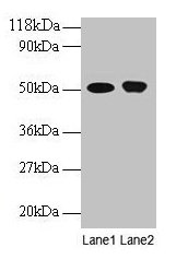
Figure 1. Western blot analysis of ODC1 using anti-ODC1 antibody (A03138-1). Electrophoresis was performed on a 5-20% SDS-PAGE gel at 70V (Stacking gel) / 90V (Resolving gel) for 2-3 hours. The sample well of each lane was loaded with 30 ug of sample under reducing conditions. Lane 1: human Hela whole cell lysates, Lane 2: human HepG2 whole cell lysates, Lane 3: human LNCAP whole cell lysates, Lane 4: human 293T whole cell lysates, Lane 5: rat thymus tissue lysates, Lane 6: rat RH35 whole cell lysates, Lane 7: mouse NIH/3T3 whole cell lysates. After electrophoresis, proteins were transferred to a nitrocellulose membrane at 150 mA for 50-90 minutes. Blocked the membrane with 5% non-fat milk/TBS for 1.5 hour at RT. The membrane was incubated with rabbit anti-ODC1 antigen affinity purified polyclonal antibody (Catalog # A03138-1) at 0.5 microg/mL overnight at 4°C, then washed with TBS-0.1%Tween 3 times with 5 minutes each and probed with a goat anti-rabbit IgG-HRP secondary antibody at a dilution of 1:5000 for 1.5 hour at RT. The signal is developed using an Enhanced Chemiluminescent detection (ECL) kit (Catalog # EK1002) with Tanon 5200 system. A specific band was detected for ODC1 at approximately 51 kDa. The expected band size for ODC1 is at 51,55 kDa.
Figure 1. Western blot analysis of ODC1 using anti-ODC1 antibody (A03138-1). Electrophoresis was performed on a 5-20% SDS-PAGE gel at 70V (Stacking gel) / 90V (Resolving gel) for 2-3 hours. The sample well of each lane was loaded with 30 ug of sample under reducing conditions. Lane 1: human Hela whole cell lysates, Lane 2: human HepG2 whole cell lysates, Lane 3: human LNCAP whole cell lysates, Lane 4: human 293T whole cell lysates, Lane 5: rat thymus tissue lysates, Lane 6: rat RH35 whole cell lysates, Lane 7: mouse NIH/3T3 whole cell lysates. After electrophoresis, proteins were transferred to a nitrocellulose membrane at 150 mA for 50-90 minutes. Blocked the membrane with 5% non-fat milk/TBS for 1.5 hour at RT. The membrane was incubated with rabbit anti-ODC1 antigen affinity purified polyclonal antibody (Catalog # A03138-1) at 0.5 microg/mL overnight at 4°C, then washed with TBS-0.1%Tween 3 times with 5 minutes each and probed with a goat anti-rabbit IgG-HRP secondary antibody at a dilution of 1:5000 for 1.5 hour at RT. The signal is developed using an Enhanced Chemiluminescent detection (ECL) kit (Catalog # EK1002) with Tanon 5200 system. A specific band was detected for ODC1 at approximately 51 kDa. The expected band size for ODC1 is at 51,55 kDa.
Anti-ODC1 Antibody Picoband(r)
A03138-1-CARRIER-FREE
ApplicationsWestern Blot, ELISA
Product group Antibodies
ReactivityHuman, Mouse, Rat
TargetODC1
Overview
- SupplierBoster Bio
- Product NameAnti-ODC1 Antibody Picoband(r)
- Delivery Days Customer9
- ApplicationsWestern Blot, ELISA
- CertificationResearch Use Only
- ClonalityPolyclonal
- Concentration500 ug/ml
- Gene ID4953
- Target nameODC1
- Target descriptionornithine decarboxylase 1
- Target synonymsBABS, NEDBA, NEDBIA, ODC, ornithine decarboxylase
- HostRabbit
- Protein IDP11926
- Protein NameOrnithine decarboxylase
- Scientific DescriptionBoster Bio Anti-ODC1 Antibody Picoband® catalog # A03138-1. Tested in WB, ELISA applications. This antibody reacts with Human, Mouse, Rat. The brand Picoband indicates this is a premium antibody that guarantees superior quality, high affinity, and strong signals with minimal background in Western blot applications. Only our best-performing antibodies are designated as Picoband, ensuring unmatched performance.
- ReactivityHuman, Mouse, Rat
- Storage Instruction-20°C,2°C to 8°C
- UNSPSC12352203
Related products
Product group Antibodies
Anti-ODC1 Antibody144-01948
ApplicationsWestern Blot, ImmunoHistoChemistry
ReactivityHuman, Mouse, Rat
TargetODC1
- SizePrice
Product group Antibodies

Anti-ODC1 [ODC1-2]AB03819-10.0
ApplicationsWestern Blot, ELISA, ImmunoHistoChemistry
ReactivityHuman
TargetODC1
- SizePrice
Product group Antibodies
Anti-ODC1 AntibodyA30530
ApplicationsWestern Blot, ImmunoHistoChemistry
ReactivityHuman, Mouse, Rat
- SizePrice
Product group Antibodies
References
ODC1 Polyclonal AntibodyBS-1294R
ApplicationsImmunoFluorescence, Western Blot, ELISA, ImmunoCytoChemistry, ImmunoHistoChemistry, ImmunoHistoChemistry Frozen, ImmunoHistoChemistry Paraffin
ReactivityBovine, Canine, Equine, Human, Mouse, Porcine, Rat
TargetODC1
- SizePrice
Product group Antibodies
Odc1 Polyclonal AntibodyCAC07342
ApplicationsImmunoFluorescence, Western Blot, ELISA, ImmunoHistoChemistry
TargetODC1
- SizePrice
Product group Antibodies
ODC1 AntibodyCSB-PA00535A0RB
ApplicationsImmunoFluorescence, Western Blot, ELISA, ImmunoHistoChemistry
ReactivityHuman
TargetODC1
- SizePrice
![Various whole cell extracts (30 μg) were separated by 10% SDS-PAGE, and the membrane was blotted with ODC antibody [N3C3] (GTX101521) diluted at 1:1000. The HRP-conjugated anti-rabbit IgG antibody (GTX213110-01) was used to detect the primary antibody, and the signal was developed with Trident ECL plus-Enhanced. Corresponding RNA expression data for the same cell lines are based on Human Protein Atlas program.](https://www.genetex.com/upload/website/prouct_img/normal/GTX101521/GTX101521_44804_20221202_WB_TPM_watermark_22122722_565.webp)
Product group Antibodies
ODC antibody [N3C3]GTX101521
ApplicationsWestern Blot, ImmunoHistoChemistry, ImmunoHistoChemistry Paraffin
ReactivityHuman, Mouse, Rat
TargetODC1
- SizePrice
Product group Antibodies

ODC1 / Ornithine Decarboxylase AntibodyLS-C331782
ApplicationsWestern Blot, ImmunoHistoChemistry
ReactivityHuman, Mouse
TargetODC1
- SizePrice