


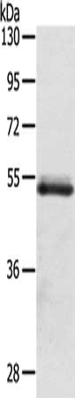
Figure 1. Western blot analysis of OLMF4 using anti-OLMF4 antibody (A04094-1). Electrophoresis was performed on a 5-20% SDS-PAGE gel at 70V (Stacking gel) / 90V (Resolving gel) for 2-3 hours. The sample well of each lane was loaded with 50ug of sample under reducing conditions. Lane 1: human placenta tissue lysates Lane 2: human U2OS whole cell lysates Lane 3: human U-87MG whole cell lysates Lane 4: human HL-60 whole cell lysates Lane 5: human K562 whole cell lysates Lane 6: human THP-1 whole cell lysates After Electrophoresis, proteins were transferred to a Nitrocellulose membrane at 150mA for 50-90 minutes. Blocked the membrane with 5% Non-fat Milk/ TBS for 1.5 hour at RT. The membrane was incubated with rabbit anti-OLMF4 antigen affinity purified polyclonal antibody (Catalog # A04094-1) at 0.5 microg/mL overnight at 4°C, then washed with TBS-0.1%Tween 3 times with 5 minutes each and probed with a goat anti-rabbit IgG-HRP secondary antibody at a dilution of 1:10000 for 1.5 hour at RT. The signal is developed using an Enhanced Chemiluminescent detection (ECL) kit (Catalog # EK1002) with Tanon 5200 system. A specific band was detected for OLMF4 at approximately 75KD. The expected band size for OLMF4 is at 57KD.
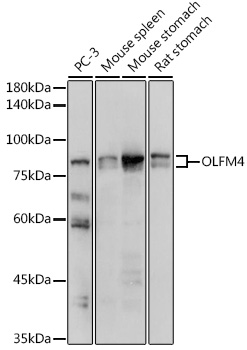
. Electrophoresis was performed on a 5-20% SDS-PAGE gel at 70V (Stacking gel) / 90V (Resolving gel) for 2-3 hours. The sample well of each lane was loaded with 50ug of sample under reducing conditions. Lane 1: rat spleen tissue lysates Lane 2: rat kidney tissue lysates Lane 3: mouse kidney tissue lysates Lane 4: mouse lung tissue lysates Lane 5: mouse SP20 whole cell lysates After Electrophoresis, proteins were transferred to a Nitrocellulose membrane at 150mA for 50-90 minutes. Blocked the membrane with 5% Non-fat Milk/ TBS for 1.5 hour at RT. The membrane was incubated with rabbit anti-OLMF4 antigen affinity purified polyclonal antibody (Catalog # A04094-1) at 0.5 microg/mL overnight at 4°C, then washed with TBS-0.1%Tween 3 times with 5 minutes each and probed with a goat anti-rabbit IgG-HRP secondary antibody at a dilution of 1:10000 for 1.5 hour at RT. The signal is developed using an Enhanced Chemiluminescent detection (ECL) kit (Catalog # EK1002) with Tanon 5200 system. A specific band was detected for OLMF4 at approximately 75KD. The expected band size for OLMF4 is at 57KD.")
. OLMF4 was detected in paraffin-embedded section of human colon cancer tissues. Heat mediated antigen retrieval was performed in citrate buffer (pH6, epitope retrieval solution) for 20 mins. The tissue section was blocked with 10% goat serum. The tissue section was then incubated with 1microg/ml rabbit anti-OLMF4 Antibody (A04094-1) overnight at 4°C. Biotinylated goat anti-rabbit IgG was used as secondary antibody and incubated for 30 minutes at 37°C. The tissue section was developed using Strepavidin-Biotin-Complex (SABC)(Catalog # SA1022) with DAB as the chromogen.")
. OLMF4 was detected in paraffin-embedded section of human rectal cancer tissues. Heat mediated antigen retrieval was performed in citrate buffer (pH6, epitope retrieval solution) for 20 mins. The tissue section was blocked with 10% goat serum. The tissue section was then incubated with 1microg/ml rabbit anti-OLMF4 Antibody (A04094-1) overnight at 4°C. Biotinylated goat anti-rabbit IgG was used as secondary antibody and incubated for 30 minutes at 37°C. The tissue section was developed using Strepavidin-Biotin-Complex (SABC)(Catalog # SA1022) with DAB as the chromogen.")
Figure 1. Western blot analysis of OLMF4 using anti-OLMF4 antibody (A04094-1). Electrophoresis was performed on a 5-20% SDS-PAGE gel at 70V (Stacking gel) / 90V (Resolving gel) for 2-3 hours. The sample well of each lane was loaded with 50ug of sample under reducing conditions. Lane 1: human placenta tissue lysates Lane 2: human U2OS whole cell lysates Lane 3: human U-87MG whole cell lysates Lane 4: human HL-60 whole cell lysates Lane 5: human K562 whole cell lysates Lane 6: human THP-1 whole cell lysates After Electrophoresis, proteins were transferred to a Nitrocellulose membrane at 150mA for 50-90 minutes. Blocked the membrane with 5% Non-fat Milk/ TBS for 1.5 hour at RT. The membrane was incubated with rabbit anti-OLMF4 antigen affinity purified polyclonal antibody (Catalog # A04094-1) at 0.5 microg/mL overnight at 4°C, then washed with TBS-0.1%Tween 3 times with 5 minutes each and probed with a goat anti-rabbit IgG-HRP secondary antibody at a dilution of 1:10000 for 1.5 hour at RT. The signal is developed using an Enhanced Chemiluminescent detection (ECL) kit (Catalog # EK1002) with Tanon 5200 system. A specific band was detected for OLMF4 at approximately 75KD. The expected band size for OLMF4 is at 57KD.
Anti-OLFM4 Antibody Picoband(r)
A04094-1-CARRIER-FREE
ApplicationsWestern Blot, ELISA, ImmunoHistoChemistry
Product group Antibodies
ReactivityHuman, Mouse, Rat
TargetOLFM4
Overview
- SupplierBoster Bio
- Product NameAnti-OLFM4 Antibody Picoband(r)
- Delivery Days Customer9
- ApplicationsWestern Blot, ELISA, ImmunoHistoChemistry
- CertificationResearch Use Only
- ClonalityPolyclonal
- Concentration500 ug/ml
- Gene ID10562
- Target nameOLFM4
- Target descriptionolfactomedin 4
- Target synonymsGC1, GW112, OLM4, OlfD, UNQ362, bA209J19.1, hGC-1, hOLfD, pDP4, olfactomedin-4, G-CSF-stimulated clone 1 protein, antiapoptotic protein GW112, olfactoimedin, tiarin
- HostRabbit
- IsotypeIgG
- Protein IDQ6UX06
- Protein NameOlfactomedin-4
- Scientific DescriptionBoster Bio Anti-OLFM4 Antibody Picoband® catalog # A04094-1. Tested in ELISA, IHC, WB applications. This antibody reacts with Human, Mouse, Rat. The brand Picoband indicates this is a premium antibody that guarantees superior quality, high affinity, and strong signals with minimal background in Western blot applications. Only our best-performing antibodies are designated as Picoband, ensuring unmatched performance.
- ReactivityHuman, Mouse, Rat
- Storage Instruction-20°C,2°C to 8°C
- UNSPSC12352203
Related products
Product group Antibodies
OLFM4 AntibodyCSB-PA252042
ApplicationsWestern Blot, ELISA
ReactivityHuman
TargetOLFM4
- SizePrice
Product group Antibodies
Anti-OLFM4 AntibodyA87732
ApplicationsImmunoFluorescence, Western Blot, ImmunoCytoChemistry
ReactivityHuman, Mouse, Rat
- SizePrice
Product group Antibodies

ApplicationsFlow Cytometry, ImmunoFluorescence, ELISA, ImmunoCytoChemistry
ReactivityHuman
TargetOLFM4
- SizePrice

Product group Antibodies
Anti-OLFM4 AntibodyHPA077718
ApplicationsImmunoHistoChemistry
ReactivityHuman
TargetOLFM4
- SizePrice
Product group Antibodies
Olfm4 Polyclonal AntibodyCAC11335
ApplicationsWestern Blot, ELISA, ImmunoHistoChemistry
ReactivityRat
TargetOLFM4
- SizePrice
Product group Antibodies
ApplicationsFlow Cytometry, Western Blot, ImmunoCytoChemistry
ReactivityHuman, Mouse, Rat
TargetOLFM4
- SizePrice
Product group Antibodies
Anti-OLFM4 Antibody144-63727
ApplicationsWestern Blot
ReactivityHuman, Mouse, Rat
TargetOLFM4
- SizePrice