

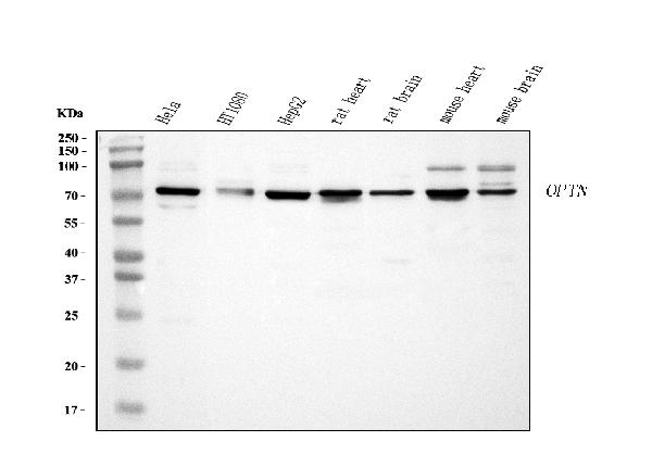
Figure 1. Western blot analysis of Optineurin using anti-Optineurin antibody (PB9343). Electrophoresis was performed on a 5-20% SDS-PAGE gel at 70V (Stacking gel) / 90V (Resolving gel) for 2-3 hours. The sample well of each lane was loaded with 30 ug of sample under reducing conditions. Lane 1: human Hela whole cell lysates, Lane 2: human HT1080 whole cell lysates, Lane 3: huamn HepG2 whole cell lysates, Lane 4: rat heart tissue lysates, Lane 5: rat brain tissue lysates, Lane 6: mouse heart tissuel lysates, Lane 7: mouse brain tissue lysates. After electrophoresis, proteins were transferred to a nitrocellulose membrane at 150 mA for 50-90 minutes. Blocked the membrane with 5% non-fat milk/TBS for 1.5 hour at RT. The membrane was incubated with rabbit anti-Optineurin antigen affinity purified polyclonal antibody (Catalog # PB9343) at 0.5 microg/mL overnight at 4°C, then washed with TBS-0.1%Tween 3 times with 5 minutes each and probed with a goat anti-rabbit IgG-HRP secondary antibody at a dilution of 1:5000 for 1.5 hour at RT. The signal is developed using an Enhanced Chemiluminescent detection (ECL) kit (Catalog # EK1002) with Tanon 5200 system. A specific band was detected for Optineurin at approximately 75 kDa. The expected band size for Optineurin is at 66 kDa.
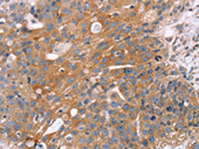
. Optineurin was detected in a paraffin-embedded section of human gall bladder adenosquamous carcinoma tissue. Heat mediated antigen retrieval was performed in EDTA buffer (pH 8.0, epitope retrieval solution). The tissue section was blocked with 10% goat serum. The tissue section was then incubated with 2 microg/ml rabbit anti-Optineurin Antibody (PB9343) overnight at 4°C. Biotinylated goat anti-rabbit IgG was used as secondary antibody and incubated for 30 minutes at 37°C. The tissue section was developed using Strepavidin-Biotin-Complex (SABC) (Catalog # SA1022) with DAB as the chromogen.")
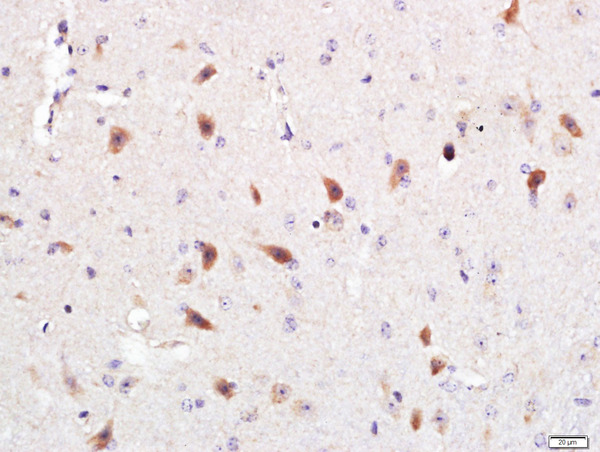
. Optineurin was detected in a paraffin-embedded section of mouse brain tissue. Heat mediated antigen retrieval was performed in EDTA buffer (pH 8.0, epitope retrieval solution). The tissue section was blocked with 10% goat serum. The tissue section was then incubated with 2 microg/ml rabbit anti-Optineurin Antibody (PB9343) overnight at 4°C. Biotinylated goat anti-rabbit IgG was used as secondary antibody and incubated for 30 minutes at 37°C. The tissue section was developed using Strepavidin-Biotin-Complex (SABC) (Catalog # SA1022) with DAB as the chromogen.")
. Optineurin was detected in a paraffin-embedded section of rat brain tissue. Heat mediated antigen retrieval was performed in EDTA buffer (pH 8.0, epitope retrieval solution). The tissue section was blocked with 10% goat serum. The tissue section was then incubated with 2 microg/ml rabbit anti-Optineurin Antibody (PB9343) overnight at 4°C. Biotinylated goat anti-rabbit IgG was used as secondary antibody and incubated for 30 minutes at 37°C. The tissue section was developed using Strepavidin-Biotin-Complex (SABC) (Catalog # SA1022) with DAB as the chromogen.")
. Overlay histogram showing A431 cells stained with PB9343 (Blue line). To facilitate intracellular staining, cells were fixed with 4% paraformaldehyde and permeabilized with permeabilization buffer. The cells were blocked with 10% normal goat serum. And then incubated with rabbit anti-Optineurin Antibody (PB9343, 1 microg/1x106 cells) for 30 min at 20°C. DyLight®488 conjugated goat anti-rabbit IgG (BA1127, 5-10 microg/1x106 cells) was used as secondary antibody for 30 minutes at 20°C. Isotype control antibody (Green line) was rabbit IgG (1 microg/1x106) used under the same conditions. Unlabelled sample without incubation with primary antibody and secondary antibody (Red line) was used as a blank control.")
Figure 1. Western blot analysis of Optineurin using anti-Optineurin antibody (PB9343). Electrophoresis was performed on a 5-20% SDS-PAGE gel at 70V (Stacking gel) / 90V (Resolving gel) for 2-3 hours. The sample well of each lane was loaded with 30 ug of sample under reducing conditions. Lane 1: human Hela whole cell lysates, Lane 2: human HT1080 whole cell lysates, Lane 3: huamn HepG2 whole cell lysates, Lane 4: rat heart tissue lysates, Lane 5: rat brain tissue lysates, Lane 6: mouse heart tissuel lysates, Lane 7: mouse brain tissue lysates. After electrophoresis, proteins were transferred to a nitrocellulose membrane at 150 mA for 50-90 minutes. Blocked the membrane with 5% non-fat milk/TBS for 1.5 hour at RT. The membrane was incubated with rabbit anti-Optineurin antigen affinity purified polyclonal antibody (Catalog # PB9343) at 0.5 microg/mL overnight at 4°C, then washed with TBS-0.1%Tween 3 times with 5 minutes each and probed with a goat anti-rabbit IgG-HRP secondary antibody at a dilution of 1:5000 for 1.5 hour at RT. The signal is developed using an Enhanced Chemiluminescent detection (ECL) kit (Catalog # EK1002) with Tanon 5200 system. A specific band was detected for Optineurin at approximately 75 kDa. The expected band size for Optineurin is at 66 kDa.
Anti-Optineurin/OPTN Antibody Picoband(r)
PB9343-CARRIER-FREE
ApplicationsFlow Cytometry, Western Blot, ImmunoHistoChemistry
Product group Antibodies
ReactivityHuman, Mouse, Rat
TargetOPTN
Overview
- SupplierBoster Bio
- Product NameAnti-Optineurin/OPTN Antibody Picoband(r)
- Delivery Days Customer9
- Application Supplier NoteTested Species: In-house tested species with positive results. Other applications have not been tested. Optimal dilutions should be determined by end users.
- ApplicationsFlow Cytometry, Western Blot, ImmunoHistoChemistry
- CertificationResearch Use Only
- ClonalityPolyclonal
- Concentration500 ug/ml
- Gene ID10133
- Target nameOPTN
- Target descriptionoptineurin
- Target synonymsALS12, FIP2, GLC1E, HIP7, HYPL, NRP, TFIIIA-INTP, optineurin, E3-14.7K-interacting protein, FIP-2, HIP-7, Huntingtin interacting protein L, huntingtin yeast partner L, huntingtin-interacting protein 7, huntingtin-interacting protein L, nemo-related protein, optic neuropathy-inducing protein, transcription factor IIIA-interacting protein, transcrption factor IIIA-interacting protein, tumor necrosis factor alpha-inducible cellular protein containing leucine zipper domains
- HostRabbit
- IsotypeIgG
- Protein IDQ96CV9
- Protein NameOptineurin
- Scientific DescriptionBoster Bio Anti-Optineurin/OPTN Antibody Picoband® catalog # PB9343. Tested in Flow Cytometry, IHC, WB applications. This antibody reacts with Human, Mouse, Rat. The brand Picoband indicates this is a premium antibody that guarantees superior quality, high affinity, and strong signals with minimal background in Western blot applications. Only our best-performing antibodies are designated as Picoband, ensuring unmatched performance.
- ReactivityHuman, Mouse, Rat
- Storage Instruction-20°C,2°C to 8°C
- UNSPSC12352203
Related products
Product group Antibodies
Anti-OPTN AntibodyA30503
ApplicationsWestern Blot, ImmunoHistoChemistry
ReactivityHuman, Mouse, Rat
- SizePrice
Product group Antibodies
Anti-OPTN Antibody144-01845
ApplicationsWestern Blot
ReactivityHuman, Mouse
TargetOPTN
- SizePrice
Product group Antibodies
Optineurin Polyclonal AntibodyBS-13658R
ApplicationsImmunoFluorescence, Western Blot, ELISA, ImmunoCytoChemistry, ImmunoHistoChemistry, ImmunoHistoChemistry Frozen, ImmunoHistoChemistry Paraffin
ReactivityMouse
TargetOPTN
- SizePrice
Product group Antibodies
OPTN AntibodyCSB-PA548038
ApplicationsELISA, ImmunoHistoChemistry
ReactivityHuman, Mouse, Rat
TargetOPTN
- SizePrice
Product group Antibodies

OPTN / Optineurin AntibodyLS-C331724
ApplicationsWestern Blot, ImmunoHistoChemistry
ReactivityHuman, Mouse
TargetOPTN
- SizePrice

Product group Antibodies
Anti-OPTN AntibodyHPA003279
ApplicationsWestern Blot, ImmunoCytoChemistry, ImmunoHistoChemistry
ReactivityHuman
TargetOPTN
- SizePrice
Product group Antibodies
Optineurin antibodyGTX105447
ApplicationsImmunoFluorescence, ImmunoPrecipitation, Western Blot, ImmunoCytoChemistry, ImmunoHistoChemistry, ImmunoHistoChemistry Paraffin
ReactivityHuman
TargetOPTN
- SizePrice
Product group Antibodies
Anti-HSPB7 AntibodyCAB18450
ApplicationsWestern Blot, ELISA, ImmunoHistoChemistry, ImmunoHistoChemistry Paraffin
ReactivityHuman
TargetOPTN
- SizePrice