


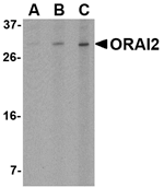
Figure 1. Western blot analysis of Orai2 using anti-Orai2 antibody (A07411-1). Electrophoresis was performed on a 5-20% SDS-PAGE gel at 70V (Stacking gel) / 90V (Resolving gel) for 2-3 hours. The sample well of each lane was loaded with 30ug of sample under reducing conditions. Lane 1: human A375 whole cell lysates, Lane 2: human Jurkat whole cell lysates, Lane 3: human HepG2 whole cell lysates, Lane 4: human K562 whole cell lysates. After Electrophoresis, proteins were transferred to a Nitrocellulose membrane at 150mA for 50-90 minutes. Blocked the membrane with 5% Non-fat Milk/ TBS for 1.5 hour at RT. The membrane was incubated with rabbit anti-Orai2 antigen affinity purified polyclonal antibody (Catalog # A07411-1) at 0.5 microg/mL overnight at 4°C, then washed with TBS-0.1%Tween 3 times with 5 minutes each and probed with a goat anti-rabbit IgG-HRP secondary antibody at a dilution of 1:5000 for 1.5 hour at RT. The signal is developed using an Enhanced Chemiluminescent detection (ECL) kit (Catalog # EK1002) with Tanon 5200 system. A specific band was detected for Orai2 at approximately 39KD. The expected band size for Orai2 is at 39KD.
Figure 1. Western blot analysis of Orai2 using anti-Orai2 antibody (A07411-1). Electrophoresis was performed on a 5-20% SDS-PAGE gel at 70V (Stacking gel) / 90V (Resolving gel) for 2-3 hours. The sample well of each lane was loaded with 30ug of sample under reducing conditions. Lane 1: human A375 whole cell lysates, Lane 2: human Jurkat whole cell lysates, Lane 3: human HepG2 whole cell lysates, Lane 4: human K562 whole cell lysates. After Electrophoresis, proteins were transferred to a Nitrocellulose membrane at 150mA for 50-90 minutes. Blocked the membrane with 5% Non-fat Milk/ TBS for 1.5 hour at RT. The membrane was incubated with rabbit anti-Orai2 antigen affinity purified polyclonal antibody (Catalog # A07411-1) at 0.5 microg/mL overnight at 4°C, then washed with TBS-0.1%Tween 3 times with 5 minutes each and probed with a goat anti-rabbit IgG-HRP secondary antibody at a dilution of 1:5000 for 1.5 hour at RT. The signal is developed using an Enhanced Chemiluminescent detection (ECL) kit (Catalog # EK1002) with Tanon 5200 system. A specific band was detected for Orai2 at approximately 39KD. The expected band size for Orai2 is at 39KD.
Anti-Orai2 Antibody Picoband(r)
A07411-1-CARRIER-FREE
ApplicationsWestern Blot, ELISA
Product group Antibodies
ReactivityHuman
TargetORAI2
Overview
- SupplierBoster Bio
- Product NameAnti-Orai2 Antibody Picoband(r)
- Delivery Days Customer9
- ApplicationsWestern Blot, ELISA
- CertificationResearch Use Only
- ClonalityPolyclonal
- Concentration500 ug/ml
- Gene ID80228
- Target nameORAI2
- Target descriptionORAI calcium release-activated calcium modulator 2
- Target synonymsC7orf19, CBCIP2, MEM142B, TMEM142B, protein orai-2, CAP-binding protein complex interacting protein 2, H_NH0514P08.8, putative protein ORAI2-2, transmembrane protein 142B
- HostRabbit
- IsotypeIgG
- Protein IDQ96SN7
- Protein NameProtein orai-2
- Scientific DescriptionBoster Bio Anti-Orai2 Antibody Picoband® catalog # A07411-1. Tested in ELISA, WB applications. This antibody reacts with Human. The brand Picoband indicates this is a premium antibody that guarantees superior quality, high affinity, and strong signals with minimal background in Western blot applications. Only our best-performing antibodies are designated as Picoband, ensuring unmatched performance.
- ReactivityHuman
- Storage Instruction-20°C,2°C to 8°C
- UNSPSC12352203
Related products
Product group Antibodies
Anti-ORAI2 AntibodyA48804
ApplicationsWestern Blot, ELISA, ImmunoCytoChemistry
ReactivityHuman, Mouse
- SizePrice
Product group Antibodies

ORAI2 AntibodyLS-C753406
ApplicationsFlow Cytometry, ImmunoFluorescence, Western Blot, ELISA, ImmunoHistoChemistry
ReactivityHuman
TargetORAI2
- SizePrice

Product group Antibodies
Anti-ORAI2 AntibodyHPA055137
ApplicationsImmunoCytoChemistry
ReactivityHuman
TargetORAI2
- SizePrice
Product group Antibodies
ORAI2 antibodyGTX120952
ApplicationsWestern Blot, ImmunoHistoChemistry, ImmunoHistoChemistry Paraffin
ReactivityHuman, Mouse, Rat
TargetORAI2
- SizePrice
Product group Antibodies
Orai2 Polyclonal AntibodyBS-9541R
ApplicationsImmunoFluorescence, Western Blot, ImmunoHistoChemistry, ImmunoHistoChemistry Paraffin
ReactivityHuman, Mouse, Rat
TargetORAI2
- SizePrice