


Figure 1. Western blot analysis of P2RY12 using anti-P2RY12 antibody (M01136). Electrophoresis was performed on a 5-20% SDS-PAGE gel at 70V (Stacking gel) / 90V (Resolving gel) for 2-3 hours. The sample well of each lane was loaded with 30 ug of sample under reducing conditions. Lane 1: human Jurkat whole cell lysates, Lane 2: human SH-SY5Y whole cell lysates, Lane 3: human U-87 MG whole cell lysates, Lane 4: human U251 whole cell lysates, Lane 5: rat brain tissue lysates, Lane 6: rat C6 whole cell lysates, Lane 7: mouse brain tissue lysates. After electrophoresis, proteins were transferred to a nitrocellulose membrane at 150 mA for 50-90 minutes. Blocked the membrane with 5% non-fat milk/TBS for 1.5 hour at RT. The membrane was incubated with rabbit anti-P2RY12 antigen affinity purified monoclonal antibody (Catalog # M01136) at 1:500 overnight at 4°C, then washed with TBS-0.1%Tween 3 times with 5 minutes each and probed with a goat anti-rabbit IgG-HRP secondary antibody at a dilution of 1:500 for 1.5 hour at RT. The signal is developed using an Enhanced Chemiluminescent detection (ECL) kit (Catalog # EK1002) with Tanon 5200 system. A specific band was detected for P2RY12 at approximately 50 kDa. The expected band size for P2RY12 is at 39 kDa.
 analysis using the Antibody at 1:50 dilution. (wb at 1:3K dilution)")
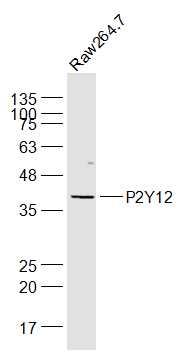
Figure 1. Western blot analysis of P2RY12 using anti-P2RY12 antibody (M01136). Electrophoresis was performed on a 5-20% SDS-PAGE gel at 70V (Stacking gel) / 90V (Resolving gel) for 2-3 hours. The sample well of each lane was loaded with 30 ug of sample under reducing conditions. Lane 1: human Jurkat whole cell lysates, Lane 2: human SH-SY5Y whole cell lysates, Lane 3: human U-87 MG whole cell lysates, Lane 4: human U251 whole cell lysates, Lane 5: rat brain tissue lysates, Lane 6: rat C6 whole cell lysates, Lane 7: mouse brain tissue lysates. After electrophoresis, proteins were transferred to a nitrocellulose membrane at 150 mA for 50-90 minutes. Blocked the membrane with 5% non-fat milk/TBS for 1.5 hour at RT. The membrane was incubated with rabbit anti-P2RY12 antigen affinity purified monoclonal antibody (Catalog # M01136) at 1:500 overnight at 4°C, then washed with TBS-0.1%Tween 3 times with 5 minutes each and probed with a goat anti-rabbit IgG-HRP secondary antibody at a dilution of 1:500 for 1.5 hour at RT. The signal is developed using an Enhanced Chemiluminescent detection (ECL) kit (Catalog # EK1002) with Tanon 5200 system. A specific band was detected for P2RY12 at approximately 50 kDa. The expected band size for P2RY12 is at 39 kDa.
Anti-P2Y12 Rabbit Monoclonal Antibody
M01136
ApplicationsImmunoPrecipitation, Western Blot
Product group Antibodies
ReactivityHuman, Mouse, Rat
TargetP2RY12
Overview
- SupplierBoster Bio
- Product NameAnti-P2Y12 Rabbit Monoclonal Antibody
- Delivery Days Customer9
- ApplicationsImmunoPrecipitation, Western Blot
- CertificationResearch Use Only
- ClonalityMonoclonal
- Clone ID28P17
- Gene ID64805
- Target nameP2RY12
- Target descriptionpurinergic receptor P2Y12
- Target synonymsADPG-R, BDPLT8, HORK3, P2T(AC), P2Y(12)R, P2Y(AC), P2Y(ADP), P2Y(cyc), P2Y12, SP1999, P2Y purinoceptor 12, ADP-glucose receptor, G-protein coupled receptor SP1999, Gi-coupled ADP receptor HORK3, P2Y12 platelet ADP receptor, purinergic receptor P2RY12, purinergic receptor P2Y, G-protein coupled, 12, putative G-protein coupled receptor
- HostRabbit
- IsotypeIgG
- Protein IDQ9H244
- Protein NameP2Y purinoceptor 12
- Scientific DescriptionBoster Bio Anti-P2Y12 Rabbit Monoclonal Antibody catalog # M01136. Tested in WB, IP applications. This antibody reacts with Human, Mouse, Rat.
- ReactivityHuman, Mouse, Rat
- Storage Instruction-20°C
- UNSPSC12352203
Related products
Product group Antibodies
Anti-P2RY12 Antibody144-61316
ApplicationsWestern Blot
ReactivityHuman, Mouse, Rat
TargetP2RY12
- SizePrice
Product group Antibodies

P2RY12 / P2Y12 AntibodyLS-C746792
ApplicationsWestern Blot
ReactivityHuman, Mouse, Rat
TargetP2RY12
- SizePrice

Product group Antibodies
Anti-P2Y12 AntibodyA90184
ApplicationsWestern Blot
ReactivityHuman, Mouse, Rat
- SizePrice
Product group Antibodies
P2Y12 Polyclonal AntibodyBS-12072R
ApplicationsImmunoFluorescence, Western Blot, ELISA, ImmunoCytoChemistry
ReactivityBovine, Canine, Equine, Human, Mouse, Porcine, Rabbit, Rat, Sheep
TargetP2RY12
- SizePrice
![P2Y12 antibody [HL2829] detects P2Y12 protein at cell membrane by immunofluorescent analysis. Sample: THP-1 cells were fixed in 4% paraformaldehyde at RT for 15 min. Green: P2Y12 stained by P2Y12 antibody [HL2829] (GTX640100) diluted at 1:500. Blue: Fluoroshield with DAPI (GTX30920).](https://www.genetex.com/upload/website/prouct_img/normal/GTX640100/GTX640100_T-45355_20240426_ICC_IF_24051400_495.webp)
Product group Antibodies
P2Y12 antibody [HL2829]GTX640100
ApplicationsImmunoFluorescence, ImmunoCytoChemistry
ReactivityHuman
TargetP2RY12
- SizePrice

Product group Antibodies
Anti-P2RY12 AntibodyHPA013796
ApplicationsImmunoHistoChemistry
ReactivityHuman
TargetP2RY12
- SizePrice