


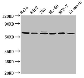
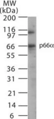
Figure 1. Western blot analysis of p66 alpha using anti-p66 alpha antibody (M09194). Electrophoresis was performed on a 5-20% SDS-PAGE gel at 70V (Stacking gel) / 90V (Resolving gel) for 2-3 hours. The sample well of each lane was loaded with 30 ug of sample under reducing conditions. Lane 1: human K562 whole cell lysates, Lane 2: human Daudi whole cell lysates, Lane 3: human HL-60 whole cell lysates, Lane 7: mouse thymus tissue lysates. After electrophoresis, proteins were transferred to a nitrocellulose membrane at 150 mA for 50-90 minutes. Blocked the membrane with 5% non-fat milk/TBS for 1.5 hour at RT. The membrane was incubated with mouse anti-p66 alpha antigen affinity purified monoclonal antibody (Catalog # M09194) at 1:500 overnight at 4°C, then washed with TBS-0.1%Tween 3 times with 5 minutes each and probed with a goat anti-mouse IgG-HRP secondary antibody at a dilution of 1:10000 for 1.5 hour at RT. The signal is developed using an Enhanced Chemiluminescent detection (ECL) kit (Catalog # EK1002) with Tanon 5200 system. A specific band was detected for p66 alpha at approximately 68 kDa. The expected band size for p66 alpha is at 68 kDa.
Figure 1. Western blot analysis of p66 alpha using anti-p66 alpha antibody (M09194). Electrophoresis was performed on a 5-20% SDS-PAGE gel at 70V (Stacking gel) / 90V (Resolving gel) for 2-3 hours. The sample well of each lane was loaded with 30 ug of sample under reducing conditions. Lane 1: human K562 whole cell lysates, Lane 2: human Daudi whole cell lysates, Lane 3: human HL-60 whole cell lysates, Lane 7: mouse thymus tissue lysates. After electrophoresis, proteins were transferred to a nitrocellulose membrane at 150 mA for 50-90 minutes. Blocked the membrane with 5% non-fat milk/TBS for 1.5 hour at RT. The membrane was incubated with mouse anti-p66 alpha antigen affinity purified monoclonal antibody (Catalog # M09194) at 1:500 overnight at 4°C, then washed with TBS-0.1%Tween 3 times with 5 minutes each and probed with a goat anti-mouse IgG-HRP secondary antibody at a dilution of 1:10000 for 1.5 hour at RT. The signal is developed using an Enhanced Chemiluminescent detection (ECL) kit (Catalog # EK1002) with Tanon 5200 system. A specific band was detected for p66 alpha at approximately 68 kDa. The expected band size for p66 alpha is at 68 kDa.
Anti-p66 alpha Rabbit Monoclonal Antibody
M09194
ApplicationsFlow Cytometry, ImmunoFluorescence, Western Blot, ImmunoCytoChemistry, ImmunoHistoChemistry
Product group Antibodies
ReactivityHuman, Mouse
TargetGATAD2A
Overview
- SupplierBoster Bio
- Product NameAnti-p66 alpha Rabbit Monoclonal Antibody
- Delivery Days Customer9
- ApplicationsFlow Cytometry, ImmunoFluorescence, Western Blot, ImmunoCytoChemistry, ImmunoHistoChemistry
- CertificationResearch Use Only
- ClonalityMonoclonal
- Clone ID26G33
- Gene ID54815
- Target nameGATAD2A
- Target descriptionGATA zinc finger domain containing 2A
- Target synonymsp66alpha, transcriptional repressor p66-alpha, GATA zinc finger domain-containing protein 2A
- HostRabbit
- IsotypeIgG
- Protein IDQ86YP4
- Protein NameTranscriptional repressor p66-alpha
- Scientific DescriptionBoster Bio Anti-p66 alpha Rabbit Monoclonal Antibody catalog # M09194. Tested in WB, IHC, ICC/IF, Flow Cytometry applications. This antibody reacts with Human, Mouse.
- ReactivityHuman, Mouse
- Storage Instruction-20°C
- UNSPSC12352203
Related products

Product group Antibodies
Anti-GATAD2A AntibodyHPA006759
ApplicationsWestern Blot, ImmunoCytoChemistry, ImmunoHistoChemistry
ReactivityHuman, Mouse, Rat
TargetGATAD2A
- SizePrice
Product group Antibodies

GATAD2A AntibodyLS-C673074
ApplicationsWestern Blot, ELISA, ImmunoHistoChemistry, ImmunoHistoChemistry Paraffin
ReactivityHuman
TargetGATAD2A
- SizePrice
Product group Antibodies
GATAD2A AntibodyCSB-PA803156LA01HU
ApplicationsWestern Blot, ELISA, ImmunoHistoChemistry
ReactivityHuman, Rat
TargetGATAD2A
- SizePrice
Product group Antibodies
GATAD2A Polyclonal AntibodyCAC15322
ApplicationsWestern Blot, ELISA, ImmunoHistoChemistry
ReactivityRat
TargetGATAD2A
- SizePrice
Product group Antibodies
GATAD2A AntibodyPACO57284
ApplicationsWestern Blot, ELISA, ImmunoHistoChemistry
ReactivityHuman, Rat
TargetGATAD2A
- SizePrice
Product group Antibodies
ApplicationsFlow Cytometry, ImmunoFluorescence, Western Blot
ReactivityHuman, Mouse
TargetGATAD2A
- SizePrice
Product group Antibodies
p66 alpha antibodyGTX13714
ApplicationsWestern Blot, ImmunoHistoChemistry, ImmunoHistoChemistry Paraffin
ReactivityHuman, Mouse, Rat
- SizePrice