


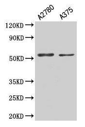
Figure 1. Western blot analysis of PAX3 using anti-PAX3 antibody (A00285-2). Electrophoresis was performed on a 5-20% SDS-PAGE gel at 70V (Stacking gel) / 90V (Resolving gel) for 2-3 hours. The sample well of each lane was loaded with 30 ug of sample under reducing conditions. Lane 1: human K562 whole cell lysates, Lane 2: human HepG2 whole cell lysates, Lane 3: human A375 whole cell lysates, Lane 4: human 293T whole cell lysates. After electrophoresis, proteins were transferred to a nitrocellulose membrane at 150 mA for 50-90 minutes. Blocked the membrane with 5% non-fat milk/TBS for 1.5 hour at RT. The membrane was incubated with rabbit anti-PAX3 antigen affinity purified polyclonal antibody (Catalog # A00285-2) at 0.5 microg/mL overnight at 4°C, then washed with TBS-0.1%Tween 3 times with 5 minutes each and probed with a goat anti-rabbit IgG-HRP secondary antibody at a dilution of 1:5000 for 1.5 hour at RT. The signal is developed using an Enhanced Chemiluminescent detection (ECL) kit (Catalog # EK1002) with Tanon 5200 system. A specific band was detected for PAX3 at approximately 69 kDa. The expected band size for PAX3 is at 53 kDa.
 and anti-Beta Tubulin antibody (M01857-3). PAX3 was detected in immunocytochemical section of A431 cell. Enzyme antigen retrieval was performed using IHC enzyme antigen retrieval reagent (AR0022) for 15 mins. The cells were blocked with 10% goat serum. And then incubated with 5 microg/mL rabbit anti-PAX3 Antibody (A00285-2) and mouse anti-Beta Tubulin antibody (M01857-3) overnight at 4°C. Cy3 Conjugated Goat Anti-Rabbit IgG (BA1032) and DyLight®488 Conjugated Goat Anti-Mouse IgG (BA1126) were used as secondary antibody at 1:500 dilution and incubated for 30 minutes at 37°C. Visualize using a fluorescence microscope and filter sets appropriate for the label used.")
Figure 1. Western blot analysis of PAX3 using anti-PAX3 antibody (A00285-2). Electrophoresis was performed on a 5-20% SDS-PAGE gel at 70V (Stacking gel) / 90V (Resolving gel) for 2-3 hours. The sample well of each lane was loaded with 30 ug of sample under reducing conditions. Lane 1: human K562 whole cell lysates, Lane 2: human HepG2 whole cell lysates, Lane 3: human A375 whole cell lysates, Lane 4: human 293T whole cell lysates. After electrophoresis, proteins were transferred to a nitrocellulose membrane at 150 mA for 50-90 minutes. Blocked the membrane with 5% non-fat milk/TBS for 1.5 hour at RT. The membrane was incubated with rabbit anti-PAX3 antigen affinity purified polyclonal antibody (Catalog # A00285-2) at 0.5 microg/mL overnight at 4°C, then washed with TBS-0.1%Tween 3 times with 5 minutes each and probed with a goat anti-rabbit IgG-HRP secondary antibody at a dilution of 1:5000 for 1.5 hour at RT. The signal is developed using an Enhanced Chemiluminescent detection (ECL) kit (Catalog # EK1002) with Tanon 5200 system. A specific band was detected for PAX3 at approximately 69 kDa. The expected band size for PAX3 is at 53 kDa.
Anti-PAX3 Antibody Picoband(r)
A00285-2-BIOTIN
ApplicationsImmunoFluorescence, Western Blot, ELISA, ImmunoCytoChemistry
Product group Antibodies
ReactivityHuman
TargetPAX3
Overview
- SupplierBoster Bio
- Product NameAnti-PAX3 Antibody Picoband(r)
- Delivery Days Customer9
- ApplicationsImmunoFluorescence, Western Blot, ELISA, ImmunoCytoChemistry
- CertificationResearch Use Only
- ClonalityPolyclonal
- Concentration500 ug/ml
- ConjugateBiotin
- Gene ID5077
- Target namePAX3
- Target descriptionpaired box 3
- Target synonymsCDHS, HUP2, PAX-3, WS1, WS3, paired box protein Pax-3, paired box homeotic gene 3, paired domain gene 3, paired domain gene HuP2, transcriptional factor PAX3
- HostRabbit
- IsotypeIgG
- Protein IDP23760
- Protein NamePaired box protein Pax-3
- Scientific DescriptionBoster Bio Anti-PAX3 Antibody Picoband® catalog # A03534-1. Tested in WB, ICC/IF, ELISA applications. This antibody reacts with Human. The brand Picoband indicates this is a premium antibody that guarantees superior quality, high affinity, and strong signals with minimal background in Western blot applications. Only our best-performing antibodies are designated as Picoband, ensuring unmatched performance.
- ReactivityHuman
- Storage Instruction-20°C,2°C to 8°C
- UNSPSC12352203
Related products

Product group Antibodies
Anti-PAX3 Antibody Picoband(r)A00285-1-CARRIER-FREE
ApplicationsFlow Cytometry, Western Blot, ELISA
ReactivityHuman, Mouse, Rat
TargetPAX3
- SizePrice
Product group Antibodies
Anti-PAX3 Antibody144-01675
ApplicationsImmunoFluorescence, Western Blot
ReactivityHuman, Mouse, Rat
TargetPAX3
- SizePrice
Product group Antibodies
PAX3 Polyclonal AntibodyCAC13856
ApplicationsImmunoFluorescence, Western Blot, ELISA, ImmunoHistoChemistry
TargetPAX3
- SizePrice
Product group Antibodies
PAX3 AntibodyCSB-PA017489HA01HU
ApplicationsImmunoFluorescence, Western Blot, ELISA, ImmunoHistoChemistry
ReactivityHuman
TargetPAX3
- SizePrice

Product group Antibodies
Anti-PAX3 AntibodyHPA063659
ApplicationsImmunoCytoChemistry, ImmunoHistoChemistry
ReactivityHuman
TargetPAX3
- SizePrice
Product group Antibodies

ApplicationsWestern Blot, ELISA, ImmunoHistoChemistry
ReactivityBovine, Human, Mouse, Rat
TargetPAX3
- SizePrice
![Non-transfected (–) and transfected (+) 293T whole cell extracts (30 μg) were separated by 10% SDS-PAGE, and the membrane was blotted with PAX3 antibody [C1C3] (GTX114094) diluted at 1:5000. The HRP-conjugated anti-rabbit IgG antibody (GTX213110-01) was used to detect the primary antibody.](https://www.genetex.com/upload/website/prouct_img/normal/GTX114094/GTX114094_40611_20220916_WB_B_22092119_496.webp)
Product group Antibodies
PAX3 antibody [C1C3]GTX114094
ApplicationsWestern Blot
ReactivityHuman
TargetPAX3
- SizePrice
Product group Antibodies

PAX3 AntibodyLS-C748944
ApplicationsWestern Blot, ImmunoHistoChemistry
ReactivityHuman, Mouse, Rat
TargetPAX3
- SizePrice