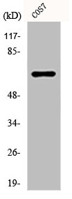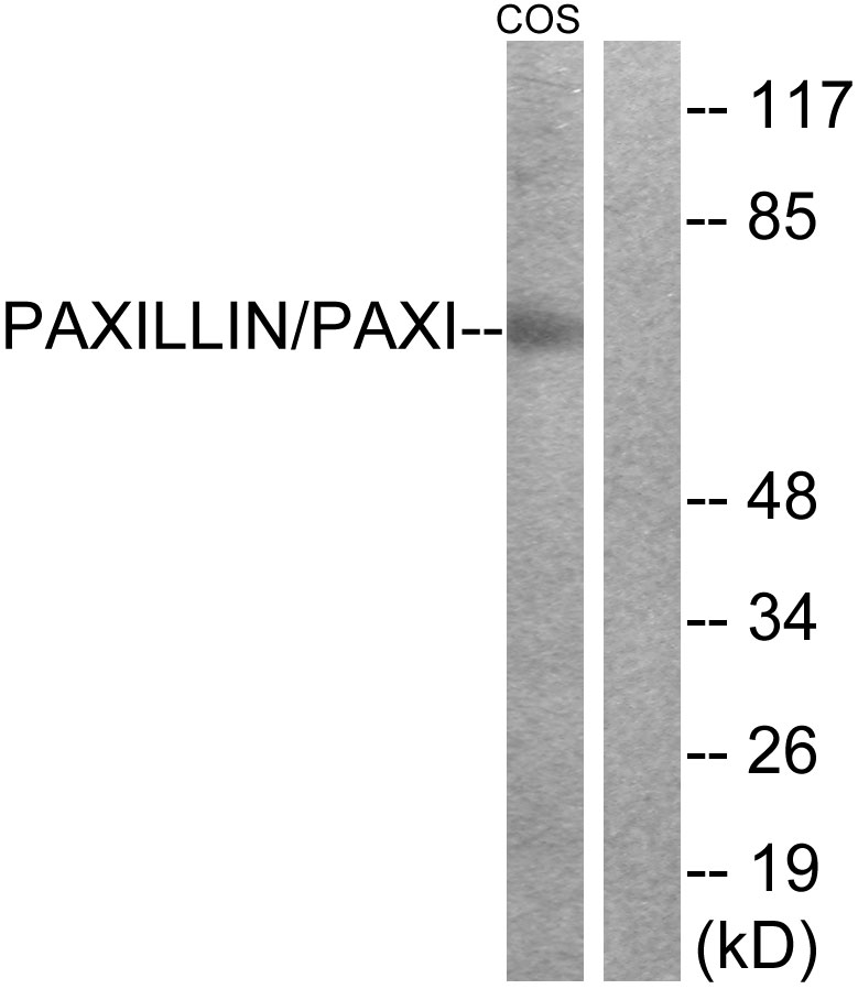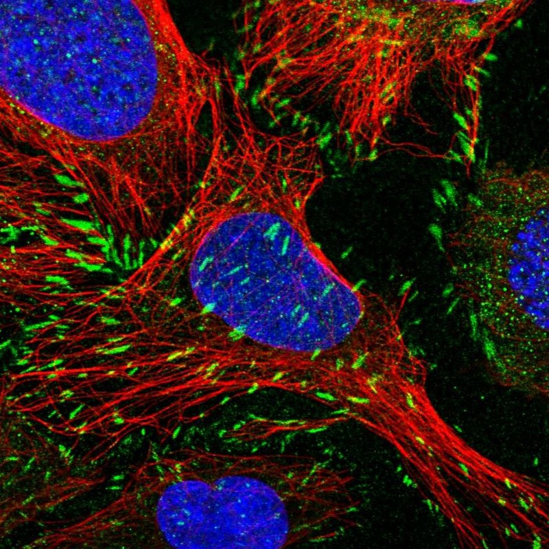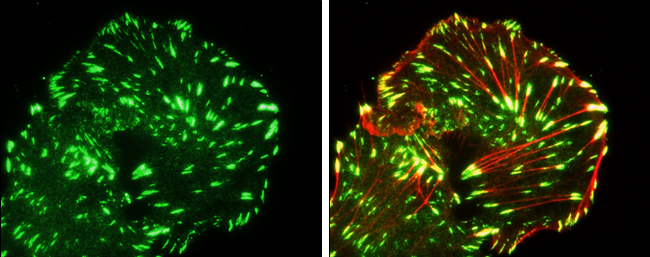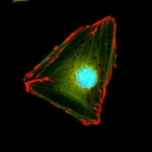
Immunocytochemical staining of HeLa cells, using anti-Paxillin rabbit monoclonal Antibody Clone RM256 (red). Actin filaments have been labeled with fluorescein phalloidin (green), and nucleus labeled with DAPI (blue).
anti-Paxillin (human), Rabbit Monoclonal (RM256)
REV-31-1137-00
ApplicationsWestern Blot, ImmunoCytoChemistry, ImmunoHistoChemistry
Product group Antibodies
ReactivityHuman
TargetPXN
Overview
- SupplierRevMAb Biosciences
- Product Nameanti-Paxillin (human), Rabbit Monoclonal (RM256)
- Delivery Days Customer2
- ApplicationsWestern Blot, ImmunoCytoChemistry, ImmunoHistoChemistry
- CertificationResearch Use Only
- ClonalityMonoclonal
- Clone IDRM256
- Gene ID5829
- Target namePXN
- Target descriptionpaxillin
- Target synonymspaxillin, testicular tissue protein Li 134
- HostRabbit
- IsotypeIgG
- Protein IDP49023
- Protein NamePaxillin
- Scientific DescriptionPaxillin is a signal transduction adaptor or scaffold protein. This class of molecule provides a structural framework to facilitate the concurrent binding of protein components in particular signalling pathways, thereby promoting efficient and sequential activation of the individual components, as well as controlling crosstalk between pathways and retaining signalling specificity. The C-terminal region of paxillin contains four LIM domains that target paxillin to focal adhesions. It is presumed through a direct association with the cytoplasmic tail of beta-integrin. The N-terminal region of paxillin is rich in protein-protein interaction sites. The proteins that bind to paxillin are diverse and include protein tyrosine kinases, such as Src and focal adhesion kinase (FAK), structural proteins, such as vinculin and actopaxin, and regulators of actin organization, such as COOL/PIX and PKL/GIT. Paxillin is tyrosine-phosphorylated by FAK and Src upon integrin engagement or growth factor stimulation, creating binding sites for the adapter protein Crk. Paxillin is expressed at focal adhesions of non-striated cells and at costameres of striated muscle cells, and it functions to adhere cells to the extracellular matrix. Paxillin has been shown to have a clinically-significant role in patients with several cancer types. Enhanced expression of paxillin has been detected in premalignant areas of hyperplasia, squamous metaplasia and goblet cell metaplasia, as well as dysplastic lesions and carcinoma in high-risk patients with lung adenocarcinoma. Mutations in PXN have been associated with enhanced tumor growth, cell proliferation and invasion in lung cancer tissues. - Recombinant Antibody. This antibody reacts to human Paxillin. This antibody may also react to mouse or rat Paxillin, as predicted by immunogen homology. Applications: WB, IHC, ICC. Source: Rabbit. Liquid. 50% Glycerol/PBS with 1% BSA and 0.09% sodium azide. Paxillin is a signal transduction adaptor or scaffold protein. This class of molecule provides a structural framework to facilitate the concurrent binding of protein components in particular signalling pathways, thereby promoting efficient and sequential activation of the individual components, as well as controlling crosstalk between pathways and retaining signalling specificity. The C-terminal region of paxillin contains four LIM domains that target paxillin to focal adhesions. It is presumed through a direct association with the cytoplasmic tail of beta-integrin. The N-terminal region of paxillin is rich in protein-protein interaction sites. The proteins that bind to paxillin are diverse and include protein tyrosine kinases, such as Src and focal adhesion kinase (FAK), structural proteins, such as vinculin and actopaxin, and regulators of actin organization, such as COOL/PIX and PKL/GIT. Paxillin is tyrosine-phosphorylated by FAK and Src upon integrin engagement or growth factor stimulation, creating binding sites for the adapter protein Crk. Paxillin is expressed at focal adhesions of non-striated cells and at costameres of striated muscle cells, and it functions to adhere cells to the extracellular matrix. Paxillin has been shown to have a clinically-significant role in patients with several cancer types. Enhanced expression of paxillin has been detected in premalignant areas of hyperplasia, squamous metaplasia and goblet cell metaplasia, as well as dysplastic lesions and carcinoma in high-risk patients with lung adenocarcinoma. Mutations in PXN have been associated with enhanced tumor growth, cell proliferation and invasion in lung cancer tissues.
- ReactivityHuman
- Storage Instruction-20°C,2°C to 8°C
- UNSPSC41116161

