Bio-Connect
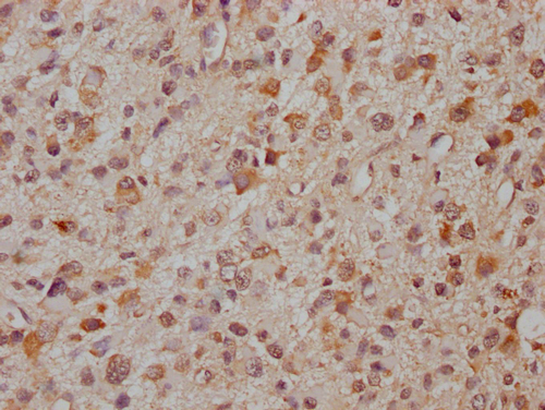
Immunohistochemical staining of human testis shows strong cytoplasmic positivity in cells in seminiferus ducts.

Anti-PCDHA8 Antibody

HPA044585
Atlas Antibodies
ApplicationsImmunoHistoChemistry
Product group Antibodies
ReactivityHuman
TargetPCDHA8
Sign in to order and to see your custom pricing.
Large volume orders?
Order with a bulk request

Overview

  • Supplier
    Atlas Antibodies
  • Product Name
    Anti-PCDHA8
  • Delivery Days Customer
    4
  • Applications
    ImmunoHistoChemistry
  • Certification
    Research Use Only
  • Clonality
    Polyclonal
  • Conjugate
    Unconjugated
  • Gene ID56140
  • Target name
    PCDHA8
  • Target description
    protocadherin alpha 8
  • Target synonyms
    KIAA0345-like 6; PCDH-ALPHA8; PCDH-alpha-8; protocadherin alpha-8
  • Host
    Rabbit
  • Isotype
    IgG
  • Protein IDQ9Y5H6
  • Protein Name
    Protocadherin alpha-8
  • Scientific Description
    Recombinant Protein Epitope Signature Tag (PrEST) antigen sequence
  • Reactivity
    Human
  • Storage Instruction
    -20°C,2°C to 8°C
  • UNSPSC
    41116161

Related products

Product group Antibodies
LifeSpan BioSciences
ApplicationsImmunoFluorescence, Western Blot, ImmunoHistoChemistry, ImmunoHistoChemistry Paraffin
TargetPCDHA8
  • SizePrice
IHC image of CSB-PA897294LA01HU diluted at 1:300 and staining in paraffin-embedded human glioma cancer performed on a Leica BondTM system. After dewaxing and hydration, antigen retrieval was mediated by high pressure in a citrate buffer (pH 6.0). Section was blocked with 10% normal goat serum 30min at RT. Then primary antibody (1% BSA) was incubated at 4°C overnight. The primary is detected by a Goat anti-rabbit IgG labeled by HRP and visualized using 0.05% DAB.
Product group Antibodies
Cusabio
PCDHA8 AntibodyCSB-PA897294LA01HU
ApplicationsELISA, ImmunoHistoChemistry
ReactivityHuman
TargetPCDHA8
  • SizePrice
Figure 1. Western blot analysis of PCDHA8 using anti-PCDHA8 antibody (A15533). Electrophoresis was performed on a 5-20% SDS-PAGE gel at 70V (Stacking gel) / 90V (Resolving gel) for 2-3 hours. The sample well of each lane was loaded with 30 ug of sample under reducing conditions. Lane 1: human PC-3 whole cell lysates, Lane 2: rat C6 whole cell lysates. After electrophoresis, proteins were transferred to a nitrocellulose membrane at 150 mA for 50-90 minutes. Blocked the membrane with 5% non-fat milk/TBS for 1.5 hour at RT. The membrane was incubated with rabbit anti-PCDHA8 antigen affinity purified polyclonal antibody (Catalog # A15533) at 0.5 microg/mL overnight at 4°C, then washed with TBS-0.1%Tween 3 times with 5 minutes each and probed with a goat anti-rabbit IgG-HRP secondary antibody at a dilution of 1:5000 for 1.5 hour at RT. The signal is developed using an Enhanced Chemiluminescent detection (ECL) kit (Catalog # EK1002) with Tanon 5200 system. A specific band was detected for PCDHA8 at approximately 150 kDa. The expected band size for PCDHA8 is at 103 kDa.
Product group Antibodies
Boster Bio
ApplicationsFlow Cytometry, Western Blot, ELISA
TargetPCDHA8
  • SizePrice
Atlas Antibodies
Protein IDQ9Y5H6
  • SizePrice
Product group Antibodies
Assay Genie
ApplicationsELISA, ImmunoHistoChemistry
TargetPCDHA8
  • SizePrice