

PD-1 receptor-induced CD4 T cell activation and proliferation by PD-1 (mouse), mAb (blocking) (1H10) (AG-20B-0075).Method: Magnetic bead affinity purified CD4+ T cells from C57BL/6 mice are stimulated in vitro with PD-1 (mouse), mAb (b
PD-1 receptor-induced CD4 T cell activation and proliferation by PD-1 (mouse), mAb (blocking) (1H10) (AG-20B-0075).Method: Magnetic bead affinity purified CD4+ T cells from C57BL/6 mice are stimulated in vitro with PD-1 (mouse), mAb (b
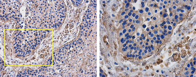
anti-PD-1 (human), mAb (AG-IHC001)
AG-20B-6020
ApplicationsELISA, ImmunoHistoChemistry
Product group Antibodies
ReactivityHuman
TargetPDCD1
Overview
- SupplierAdipoGen Life Sciences
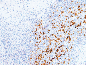
- Product Nameanti-PD-1 (human), mAb (AG-IHC001)
- Delivery Days Customer10
- ApplicationsELISA, ImmunoHistoChemistry
- CertificationResearch Use Only
- ClonalityMonoclonal
- Clone IDAG-IHC001
- Gene ID5133
- Target namePDCD1
- Target descriptionprogrammed cell death 1
- Target synonymsADMIO4, AIMTBS, CD279, PD-1, PD1, SLEB2, hPD-1, hPD-l, hSLE1, programmed cell death protein 1, programmed cell death 1 protein, protein PD-1, systemic lupus erythematosus susceptibility 2
- HostMouse
- IsotypeIgG2b
- Protein IDQ15116
- Protein NameProgrammed cell death protein 1
- Scientific DescriptionMonoclonal Antibody. Recognizes human PD-1 (CD279). Applications: ELISA, IHC. Isotype: Mouse IgG2b. Clone: AG-IHC001. Liquid. In Tris Buffer, pH 7.4, containing 1% BSA and <0.1% sodium azide. Programmed Death 1 (PD-1) is a member of the CD28/CTLA-4 family of T cell regulators, expressed as a co-receptor on the surface of activated T cells, B cells and macrophages. Several studies have suggested that the PD-1/PD-L1 signaling pathway may be linked to antitumor immunity, as PD-L1 has been shown to induce apoptosis of activated T cells or inhibit activity of cytotoxic T cells. In comparison to CD10 and Bcl-6, PD-1 is expressed by fewer B cells and has therefore been considered a more specific and useful diagnostic marker for angioimmunoblastic T cell lymphoma. Therapies targeted toward the PD-1 receptor have shown remarkable clinical responses in patients with various types of cancer, including non-small-cell lung cancer, melanoma and renal-cell cancer. This antibody is intended to qualitatively identify by light microscopy the presence of associated antigens in sections of formalin-fixed, paraffin-embedded tissue sections using IHC test methods. It has been optimized and validated using the BOND-MAX fully automated IHC&ISH stainer (see Protocol). - Programmed Death 1 (PD-1) is a member of the CD28/CTLA-4 family of T cell regulators, expressed as a co-receptor on the surface of activated T cells, B cells and macrophages. Several studies have suggested that the PD-1/PD-L1 signaling pathway may be linked to antitumor immunity, as PD-L1 has been shown to induce apoptosis of activated T cells or inhibit activity of cytotoxic T cells. In comparison to CD10 and Bcl-6, PD-1 is expressed by fewer B cells and has therefore been considered a more specific and useful diagnostic marker for angioimmunoblastic T cell lymphoma. Therapies targeted toward the PD-1 receptor have shown remarkable clinical responses in patients with various types of cancer, including non-small-cell lung cancer, melanoma and renal-cell cancer. This antibody is intended to qualitatively identify by light microscopy the presence of associated antigens in sections of formalin-fixed, paraffin-embedded tissue sections using IHC test methods. It has been optimized and validated using the BOND-MAX fully automated IHC&ISH stainer (see Protocol).
- ReactivityHuman
- Storage Instruction2°C to 8°C
- UNSPSC41116161
MSDS
Related products
Product group Antibodies
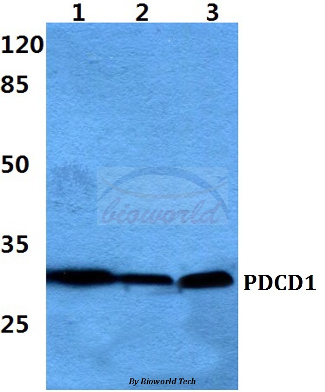
Anti-PDCD1 AntibodyA28569
ApplicationsWestern Blot
ReactivityHuman, Mouse, Rat
- SizePrice

Product group Antibodies
Anti-PDCD1 AntibodyAMAB91197
ApplicationsWestern Blot, ImmunoHistoChemistry
ReactivityHuman
TargetPDCD1
- SizePrice
Product group Antibodies

ApplicationsFlow Cytometry, ImmunoHistoChemistry
ReactivityMouse
TargetPDCD1
- SizePrice
Product group Antibodies

Anti-PD-1 [5C4.B8 (Nivolumab)]Ab00791-1.1
ApplicationsFlow Cytometry, ImmunoHistoChemistry, Neutralisation/Blocking, Other Application
ReactivityHuman, Monkey
TargetPDCD1
- SizePrice
Product group Antibodies
References
PD-1 Polyclonal AntibodyBS-1867R
ApplicationsFlow Cytometry, ImmunoFluorescence, Western Blot, ELISA, ImmunoCytoChemistry, ImmunoHistoChemistry, ImmunoHistoChemistry Frozen, ImmunoHistoChemistry Paraffin
ReactivityHuman, Mouse, Rat
TargetPDCD1
- SizePrice
Product group Antibodies
PDCD1 AntibodyCSB-PA483633
ApplicationsELISA, ImmunoHistoChemistry
ReactivityHuman
TargetPDCD1
- SizePrice
Product group Antibodies
Pdcd1 Polyclonal AntibodyCAC07144
ApplicationsImmunoFluorescence, Western Blot, ELISA, ImmunoHistoChemistry
ReactivityMouse
TargetPDCD1
- SizePrice
Product group Antibodies
PD1 antibodyGTX128435
ApplicationsWestern Blot, ImmunoHistoChemistry, ImmunoHistoChemistry Paraffin
ReactivityHuman, Mouse
TargetPDCD1
- SizePrice