

Anti-PD-1 [J43]
AB01417-2.3-VXS
ApplicationsFlow Cytometry, ImmunoPrecipitation, ELISA, ImmunoHistoChemistry, Neutralisation/Blocking, Other Application
Product group Antibodies
ReactivityMouse
TargetPdcd1
Overview
- SupplierAbsolute Antibody
- Product NameAnti-PD-1 [J43]
- Delivery Days Customer9
- Application Supplier NoteThe specificity of this antibody has been confirmed in ELISA analysis, using PD-1 extracellular domain fusion proteins (Agata et al, 1996). Additionally, in flow cytometric analysis, this antibody reacts with PD-1 cDNA-transfected BHK and CHO cells, but not with parental BHK and CHO cells, as well as reacting with lymphocytes from PD-1 cDNA transgenic mice (Agata et al, 1996). This antibody has been used to immunoprecipitate PD-1 from lysates of PD-1 cDNA-transfected BHK and CHO cells (Agata et al, 1996), in flow cytometric quantification of CD4+PD-1+ T cells in murine spleens (Kasagi et al, 2010), and in immunohistochemical analysis of acetone-fixed murine spinal cord and brain tissue sections (Salama et al, 2003). This antibody displays diverse effects in different mouse models of disease. When administered to NZB/W F1 mice, a model of lupus-like nephritis, this antibody has been shown to delay the onset of nephritis and prolong survival, through the depletion of PD-1+ T cells (Kasagi et al, 2010). Antibody-treated NZB/W F1 mice displayed decreased numbers of PD-1+ T cells, and this antibody was confirmed to trigger complement-dependent cytotoxicity in PD-1+ T cells in vitro (Kasagi et al, 2010). Conversely, administration to experimental allergic encephalitis (EAE) and NOD diabetes mice exacerbated disease, through its neutralising activity (Salama et al, 2003; Ansari et al, 2003); this antibody has been shown in vitro to inhibit binding of both PD-L1-Ig and PD-L2-Ig to PD-1 transfected BHK cells (Ansari et al, 2003).
- ApplicationsFlow Cytometry, ImmunoPrecipitation, ELISA, ImmunoHistoChemistry, Neutralisation/Blocking, Other Application
- CertificationResearch Use Only
- ClonalityMonoclonal
- Clone IDJ43
- Gene ID18566
- Target namePdcd1
- Target descriptionprogrammed cell death 1
- Target synonymsLy101, PD-1, Pdc1, programmed cell death protein 1, mPD-1, programmed cell death 1 protein, protein PD-1
- HostMouse
- IsotypeIgG2a
- Protein IDQ02242
- Protein NameProgrammed cell death protein 1
- Scientific DescriptionThis chimeric mouse antibody was made using the variable domain sequences of the original Hamster IgG format, for improved compatibility with existing reagents, assays and techniques.
- ReactivityMouse
- Storage Instruction-20°C,2°C to 8°C
- UNSPSC41116161
Related products
Product group Antibodies
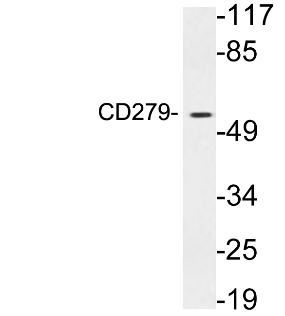
Anti-CD279 AntibodyA101172
ApplicationsWestern Blot, ELISA
ReactivityHuman
- SizePrice

Product group Antibodies
Anti-PD1/Pdcd1 Antibody Picoband(r)A00178-3-CARRIER-FREE
ApplicationsWestern Blot, ELISA, ImmunoHistoChemistry
ReactivityMouse, Rat
TargetPdcd1
- SizePrice
Product group Antibodies
PD1 antibody [MAB0839]GTX52425
ApplicationsFlow Cytometry, Neutralisation/Blocking
ReactivityMouse
TargetPdcd1
- SizePrice
Product group Antibodies
ApplicationsFlow Cytometry
ReactivityMouse
TargetPdcd1
- SizePrice