


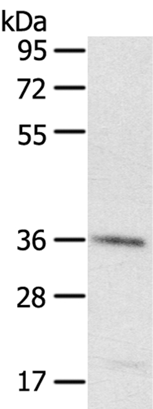
Figure 1. Western blot analysis of PDLIM4 using anti-PDLIM4 antibody (A09573-2). Electrophoresis was performed on a 5-20% SDS-PAGE gel at 70V (Stacking gel) / 90V (Resolving gel) for 2-3 hours. The sample well of each lane was loaded with 30 ug of sample under reducing conditions. Lane 1: human Hacat whole cell lysates, Lane 2: human U251 whole cell lysates, Lane 3: human A431 whole cell lysates, Lane 4: human U20S whole cell lysates. After electrophoresis, proteins were transferred to a nitrocellulose membrane at 150 mA for 50-90 minutes. Blocked the membrane with 5% non-fat milk/TBS for 1.5 hour at RT. The membrane was incubated with rabbit anti-PDLIM4 antigen affinity purified polyclonal antibody (Catalog # A09573-2) at 0.5 microg/mL overnight at 4°C, then washed with TBS-0.1%Tween 3 times with 5 minutes each and probed with a goat anti-rabbit IgG-HRP secondary antibody at a dilution of 1:5000 for 1.5 hour at RT. The signal is developed using an Enhanced Chemiluminescent detection (ECL) kit (Catalog # EK1002) with Tanon 5200 system. A specific band was detected for PDLIM4 at approximately 35 kDa. The expected band size for PDLIM4 is at 35,26 kDa.
. PDLIM4 was detected in an immunocytochemical section of U2OS cells. Enzyme antigen retrieval was performed using IHC enzyme antigen retrieval reagent (AR0022) for 15 mins. The cells were blocked with 10% goat serum. And then incubated with 5 microg/mL rabbit anti-PDLIM4 Antibody (A09573-2) overnight at 4°C. DyLight®488 Conjugated Goat Anti-Rabbit IgG (BA1127) was used as secondary antibody at 1:500 dilution and incubated for 30 minutes at 37°C. The section was counterstained with DAPI. Visualize using a fluorescence microscope and filter sets appropriate for the label used.")
. Overlay histogram showing A431 cells stained with A09573-2 (Blue line). To facilitate intracellular staining, cells were fixed with 4% paraformaldehyde and permeabilized with permeabilization buffer. The cells were blocked with 10% normal goat serum. And then incubated with rabbit anti-PDLIM4 Antibody (A09573-2, 1 microg/1x106 cells) for 30 min at 20°C. DyLight®488 conjugated goat anti-rabbit IgG (BA1127, 5-10 microg/1x106 cells) was used as secondary antibody for 30 minutes at 20°C. Isotype control antibody (Green line) was rabbit IgG (1 microg/1x106) used under the same conditions. Unlabelled sample without incubation with primary antibody and secondary antibody (Red line) was used as a blank control.")
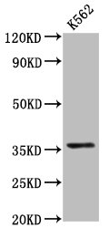
. Electrophoresis was performed on a 5-20% SDS-PAGE gel at 70V (Stacking gel) / 90V (Resolving gel) for 2-3 hours. The sample well of each lane was loaded with 30 ug of sample under reducing conditions. Lane 1: human U251 whole cell lysates, Lane 2: human U20S whole cell lysates. After electrophoresis, proteins were transferred to a nitrocellulose membrane at 150 mA for 50-90 minutes. Blocked the membrane with 5% non-fat milk/TBS for 1.5 hour at RT. The membrane was incubated with rabbit anti-PDLIM4 antigen affinity purified polyclonal antibody (A09573-2) at 0.5 microg/mL overnight at 4°C, then washed with TBS-0.1%Tween 3 times with 5 minutes each and probed with a goat anti-rabbit IgG-DyLight 647 Conjugated secondary antibody at a dilution of 1:2000 for 1.5 hour at RT. A specific band was detected for PDLIM4 at approximately 37 kDa. The expected band size for PDLIM4 is at 35 kDa.")
Figure 1. Western blot analysis of PDLIM4 using anti-PDLIM4 antibody (A09573-2). Electrophoresis was performed on a 5-20% SDS-PAGE gel at 70V (Stacking gel) / 90V (Resolving gel) for 2-3 hours. The sample well of each lane was loaded with 30 ug of sample under reducing conditions. Lane 1: human Hacat whole cell lysates, Lane 2: human U251 whole cell lysates, Lane 3: human A431 whole cell lysates, Lane 4: human U20S whole cell lysates. After electrophoresis, proteins were transferred to a nitrocellulose membrane at 150 mA for 50-90 minutes. Blocked the membrane with 5% non-fat milk/TBS for 1.5 hour at RT. The membrane was incubated with rabbit anti-PDLIM4 antigen affinity purified polyclonal antibody (Catalog # A09573-2) at 0.5 microg/mL overnight at 4°C, then washed with TBS-0.1%Tween 3 times with 5 minutes each and probed with a goat anti-rabbit IgG-HRP secondary antibody at a dilution of 1:5000 for 1.5 hour at RT. The signal is developed using an Enhanced Chemiluminescent detection (ECL) kit (Catalog # EK1002) with Tanon 5200 system. A specific band was detected for PDLIM4 at approximately 35 kDa. The expected band size for PDLIM4 is at 35,26 kDa.
Anti-PDLIM4 Antibody Picoband(r)
A09573-2-CARRIER-FREE
ApplicationsFlow Cytometry, ImmunoFluorescence, ImmunoPrecipitation, Western Blot, ELISA, ImmunoCytoChemistry
Product group Antibodies
ReactivityHuman
TargetPDLIM4
Overview
- SupplierBoster Bio
- Product NameAnti-PDLIM4 Antibody Picoband(r)
- Delivery Days Customer9
- ApplicationsFlow Cytometry, ImmunoFluorescence, ImmunoPrecipitation, Western Blot, ELISA, ImmunoCytoChemistry
- CertificationResearch Use Only
- ClonalityPolyclonal
- Concentration500 ug/ml
- Gene ID8572
- Target namePDLIM4
- Target descriptionPDZ and LIM domain 4
- Target synonymsRIL, PDZ and LIM domain protein 4, LIM domain protein, LIM protein RIL, enigma homolog, reversion-induced LIM protein
- HostRabbit
- Protein IDP50479
- Protein NamePDZ and LIM domain protein 4
- Scientific DescriptionBoster Bio Anti-PDLIM4 Antibody Picoband® catalog # A09573-2. Tested in WB, IP, ICC, IF, Flow Cytometry, ELISA applications. This antibody reacts with Human. The brand Picoband indicates this is a premium antibody that guarantees superior quality, high affinity, and strong signals with minimal background in Western blot applications. Only our best-performing antibodies are designated as Picoband, ensuring unmatched performance.
- ReactivityHuman
- Storage Instruction-20°C,2°C to 8°C
- UNSPSC12352203
Related products
Product group Antibodies
Anti-PDLIM4 AntibodyA37346
ApplicationsWestern Blot, ImmunoHistoChemistry
ReactivityHuman
- SizePrice
Product group Antibodies
RIL Polyclonal AntibodyBS-6093R
ApplicationsImmunoFluorescence, ELISA, ImmunoCytoChemistry, ImmunoHistoChemistry, ImmunoHistoChemistry Frozen, ImmunoHistoChemistry Paraffin
ReactivityBovine, Canine, Chicken, Equine, Human, Mouse, Porcine, Rabbit, Rat
TargetPDLIM4
- SizePrice
Product group Antibodies

Goat anti-PDLIM4 / RILEB06249
ApplicationsImmunoFluorescence, Western Blot, ELISA
ReactivityBovine, Human, Mouse, Rat
TargetPDLIM4
- SizePrice
Product group Antibodies
PDLIM4 Polyclonal AntibodyCAC15311
ApplicationsImmunoPrecipitation, Western Blot, ELISA
TargetPDLIM4
- SizePrice
Product group Antibodies
PDLIM4 AntibodyCSB-PA017735LA01HU
ApplicationsImmunoPrecipitation, Western Blot, ELISA
ReactivityHuman
TargetPDLIM4
- SizePrice
Product group Antibodies

PDLIM4 / RIL AntibodyLS-C401988
ApplicationsWestern Blot, ELISA, ImmunoHistoChemistry
ReactivityHuman, Mouse, Rat
TargetPDLIM4
- SizePrice

Product group Antibodies
Anti-PDLIM4-25ulHPA011912
ApplicationsWestern Blot, ImmunoCytoChemistry, ImmunoHistoChemistry
ReactivityHuman, Rat
- SizePrice
Product group Antibodies
PDLIM4 antibody [C2C3], C-termGTX106353
ApplicationsImmunoFluorescence, Western Blot, ImmunoCytoChemistry
ReactivityHuman
TargetPDLIM4
- SizePrice