


Immunohistochemical staining of human testis shows cytoplasmic positivity in cells in seminiferous ducts.
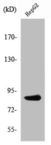
![Lane 1: Marker [kDa] 250, 130, 100, 70, 55, 35, 25, 15, 10. Lane 2: Human cell line EFO-21](https://atlasantibodies.s3.amazonaws.com/images/wb/hpa051484-wb-1.jpg "Lane 1: Marker [kDa] 250, 130, 100, 70, 55, 35, 25, 15, 10. Lane 2: Human cell line EFO-21")
Immunohistochemical staining of human testis shows cytoplasmic positivity in cells in seminiferous ducts.
Anti-PDXDC1 Antibody
HPA051484
ApplicationsWestern Blot, ImmunoCytoChemistry, ImmunoHistoChemistry
Product group Antibodies
ReactivityHuman
TargetPDXDC1
Overview
- SupplierAtlas Antibodies
- Product NameAnti-PDXDC1 Antibody
- Delivery Days Customer4
- ApplicationsWestern Blot, ImmunoCytoChemistry, ImmunoHistoChemistry
- CertificationResearch Use Only
- ClonalityPolyclonal
- ConjugateUnconjugated
- Gene ID23042
- Target namePDXDC1
- Target descriptionpyridoxal dependent decarboxylase domain containing 1
- Target synonymsLP8165, pyridoxal-dependent decarboxylase domain-containing protein 1
- HostRabbit
- IsotypeIgG
- Protein IDQ6P996
- Protein NamePyridoxal-dependent decarboxylase domain-containing protein 1
- Scientific DescriptionRecombinant Protein Epitope Signature Tag (PrEST) antigen sequence
- ReactivityHuman
- Storage Instruction-20°C,2°C to 8°C
- UNSPSC41116161
Datasheet
MSDS
Related products

Product group Antibodies
Anti-PDXDC1 AntibodyA96796
ApplicationsWestern Blot, ELISA
ReactivityHuman, Mouse, Rat
- SizePrice

Product group Antibodies
Anti-PDXDC1 Antibody Picoband(r)A11375-2-CARRIER-FREE
ApplicationsFlow Cytometry, Western Blot, ELISA, ImmunoHistoChemistry
ReactivityHuman, Mouse, Rat
TargetPDXDC1
- SizePrice
Product group Antibodies
Anti-PDXDC1 Antibody144-60825
ApplicationsWestern Blot
ReactivityHuman, Mouse, Rat
TargetPDXDC1
- SizePrice
Product group Antibodies

PDXDC1 AntibodyLS-C760992
ApplicationsWestern Blot, ImmunoHistoChemistry
ReactivityHuman
TargetPDXDC1
- SizePrice
Product group Antibodies
PDXDC1 AntibodyCSB-PA003734
ApplicationsWestern Blot, ELISA, ImmunoHistoChemistry
ReactivityHuman, Mouse, Rat
TargetPDXDC1
- SizePrice
Product group Antibodies
PDXDC1 antibodyGTX87408
ApplicationsWestern Blot
ReactivityHuman
TargetPDXDC1
- SizePrice