


Immunohistochemical staining of human kidney shows strong cytoplasmic and membranous positivity in tubular cells.
 shows similar pattern to independent antibody HPA030196 (B).")
Immunohistochemical staining of human kidney shows strong cytoplasmic and membranous positivity in tubular cells.
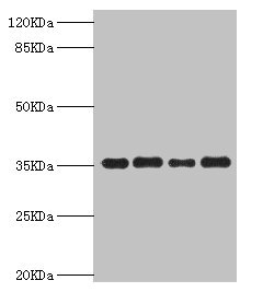
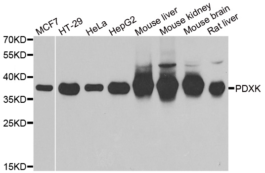
Anti-PDXK Antibody
HPA030198
ApplicationsWestern Blot, ImmunoHistoChemistry
Product group Antibodies
ReactivityHuman
TargetPDXK
Overview
- SupplierAtlas Antibodies
- Product NameAnti-PDXK Antibody
- Delivery Days Customer4
- ApplicationsWestern Blot, ImmunoHistoChemistry
- CertificationResearch Use Only
- ClonalityPolyclonal
- ConjugateUnconjugated
- Gene ID8566
- Target namePDXK
- Target descriptionpyridoxal kinase
- Target synonymsC21orf124, C21orf97, HEL-S-1a, HMSN6C, PKH, PNK, PRED79, pyridoxal kinase, epididymis secretory sperm binding protein Li 1a, pyridoxal (pyridoxine, vitamin B6) kinase, pyridoxamine kinase, pyridoxine kinase, vitamin B6 kinase
- HostRabbit
- IsotypeIgG
- Protein IDO00764
- Protein NamePyridoxal kinase
- Scientific DescriptionRecombinant Protein Epitope Signature Tag (PrEST) antigen sequence
- ReactivityHuman
- Storage Instruction-20°C,2°C to 8°C
- UNSPSC41116161
Datasheet
MSDS
Related products
Product group Antibodies
PDXK AntibodyCSB-PA017748ESR2HU
ApplicationsWestern Blot, ELISA, ImmunoHistoChemistry
ReactivityHuman, Mouse
TargetPDXK
- SizePrice

Product group Antibodies
Anti-PDXK Antibody Picoband(r)A06683-1-CARRIER-FREE
ApplicationsFlow Cytometry, Western Blot, ELISA, ImmunoHistoChemistry
ReactivityHuman, Mouse, Rat
TargetPDXK
- SizePrice
Product group Antibodies
Anti-PDXK AntibodyA31522
ApplicationsWestern Blot, ImmunoHistoChemistry
ReactivityHuman, Mouse, Rat
- SizePrice
Product group Antibodies

PDXK / PNK AntibodyLS-C830978
ApplicationsELISA, ImmunoHistoChemistry
ReactivityHuman, Mouse, Rat
TargetPDXK
- SizePrice

Product group Antibodies
Anti-PDXK AntibodyHPA030196
ApplicationsWestern Blot, ImmunoCytoChemistry, ImmunoHistoChemistry
ReactivityHuman
TargetPDXK
- SizePrice