

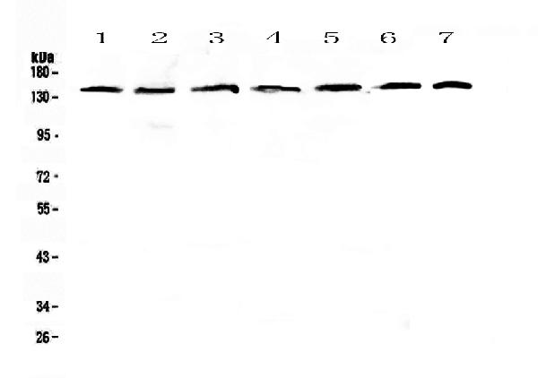
Figure 1. Western blot analysis of PERK using anti-PERK antibody (A01992-2). Electrophoresis was performed on a 5-20% SDS-PAGE gel at 70V (Stacking gel) / 90V (Resolving gel) for 2-3 hours. The sample well of each lane was loaded with 50ug of sample under reducing conditions. Lane 1: human Hela whole cell lysates, Lane 2: human COLO-320 whole cell lysates, Lane 3: human A549 whole cell lysates, Lane 4: human SK-OV-3 whole cell lysates, Lane 5: Human A431 whole cell lysates, Lane 6: rat brain tissue lysates, Lane 7: mouse brain tissue lysates. After Electrophoresis, proteins were transferred to a Nitrocellulose membrane at 150mA for 50-90 minutes. Blocked the membrane with 5% Non-fat Milk/ TBS for 1.5 hour at RT. The membrane was incubated with rabbit anti-PERK antigen affinity purified polyclonal antibody (Catalog # A01992-2) at 0.5 microg/mL overnight at 4°C, then washed with TBS-0.1%Tween 3 times with 5 minutes each and probed with a goat anti-rabbit IgG-HRP secondary antibody at a dilution of 1:10000 for 1.5 hour at RT. The signal is developed using an Enhanced Chemiluminescent detection (ECL) kit (Catalog # EK1002) with Tanon 5200 system. A specific band was detected for PERK at approximately 140KD. The expected band size for PERK is at 125KD.
. Overlay histogram showing HepG2 cells stained with A01992-2 (Blue line). To facilitate intracellular staining, cells were fixed with 4% paraformaldehyde and permeabilized with permeabilization buffer. The cells were blocked with 10% normal goat serum. And then incubated with rabbit anti-PERK Antibody (A01992-2,1microg/1x106 cells) for 30 min at 20°C. DyLight®488 conjugated goat anti-rabbit IgG (BA1127, 5-10microg/1x106 cells) was used as secondary antibody for 30 minutes at 20°C. Isotype control antibody (Green line) was rabbit IgG (1microg/1x106) used under the same conditions. Unlabelled sample without incubation with primary antibody and secondary antibody (Red line) was used as a blank control.")
Figure 1. Western blot analysis of PERK using anti-PERK antibody (A01992-2). Electrophoresis was performed on a 5-20% SDS-PAGE gel at 70V (Stacking gel) / 90V (Resolving gel) for 2-3 hours. The sample well of each lane was loaded with 50ug of sample under reducing conditions. Lane 1: human Hela whole cell lysates, Lane 2: human COLO-320 whole cell lysates, Lane 3: human A549 whole cell lysates, Lane 4: human SK-OV-3 whole cell lysates, Lane 5: Human A431 whole cell lysates, Lane 6: rat brain tissue lysates, Lane 7: mouse brain tissue lysates. After Electrophoresis, proteins were transferred to a Nitrocellulose membrane at 150mA for 50-90 minutes. Blocked the membrane with 5% Non-fat Milk/ TBS for 1.5 hour at RT. The membrane was incubated with rabbit anti-PERK antigen affinity purified polyclonal antibody (Catalog # A01992-2) at 0.5 microg/mL overnight at 4°C, then washed with TBS-0.1%Tween 3 times with 5 minutes each and probed with a goat anti-rabbit IgG-HRP secondary antibody at a dilution of 1:10000 for 1.5 hour at RT. The signal is developed using an Enhanced Chemiluminescent detection (ECL) kit (Catalog # EK1002) with Tanon 5200 system. A specific band was detected for PERK at approximately 140KD. The expected band size for PERK is at 125KD.
Anti-PERK/EIF2AK3 Antibody Picoband(r)
A01992-2-CARRIER-FREE
ApplicationsFlow Cytometry, Western Blot, ELISA
Product group Antibodies
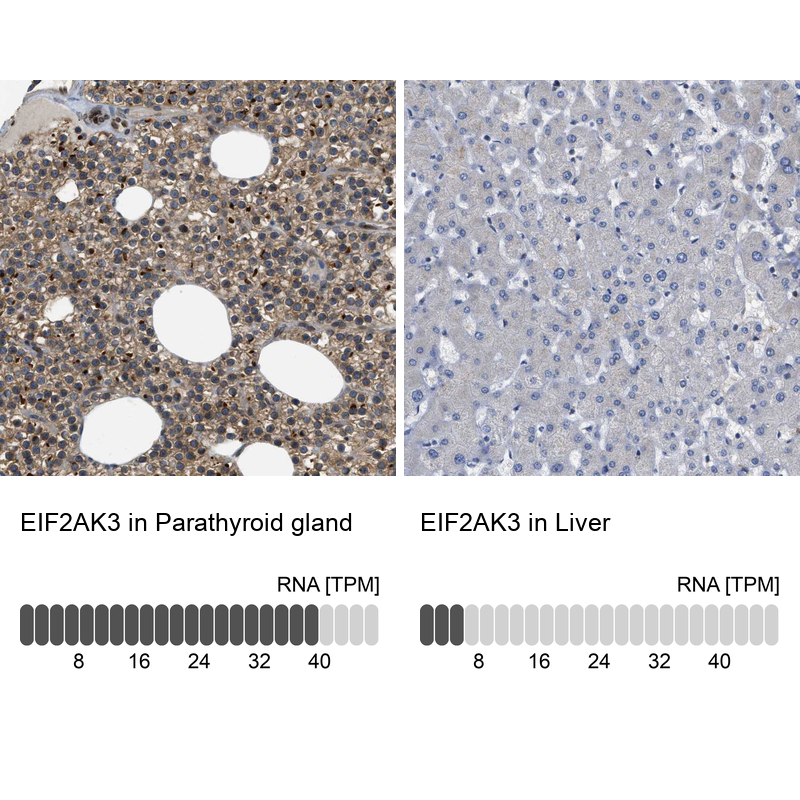
ReactivityHuman, Mouse, Rat
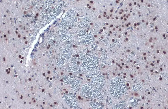
TargetEIF2AK3
Overview
- SupplierBoster Bio
- Product NameAnti-PERK/EIF2AK3 Antibody Picoband(r)
- Delivery Days Customer9
- ApplicationsFlow Cytometry, Western Blot, ELISA
- CertificationResearch Use Only
- ClonalityPolyclonal
- Concentration500 ug/ml
- Gene ID9451
- Target nameEIF2AK3
- Target descriptioneukaryotic translation initiation factor 2 alpha kinase 3
- Target synonymsPEK, PERK, WRS, eukaryotic translation initiation factor 2-alpha kinase 3, PKR-like ER kinase, PRKR-like endoplasmic reticulum kinase, pancreatic EIF2-alpha kinase, pancreatic eIF-2alpha kinase, protein tyrosine kinase EIF2AK3
- HostRabbit
- IsotypeIgG
- Protein IDQ9NZJ5
- Protein NameEukaryotic translation initiation factor 2-alpha kinase 3
- Scientific DescriptionBoster Bio Anti-PERK/EIF2AK3 Antibody Picoband® catalog # A01992-2. Tested in ELISA, Flow Cytometry, WB applications. This antibody reacts with Human, Mouse, Rat. The brand Picoband indicates this is a premium antibody that guarantees superior quality, high affinity, and strong signals with minimal background in Western blot applications. Only our best-performing antibodies are designated as Picoband, ensuring unmatched performance.
- ReactivityHuman, Mouse, Rat
- Storage Instruction-20°C,2°C to 8°C
- UNSPSC12352203
Related products
Product group Antibodies
Anti-PERK AntibodyA306749
ApplicationsImmunoFluorescence, Western Blot, ImmunoCytoChemistry
ReactivityHuman, Mouse, Rat
- SizePrice
Product group Antibodies
Anti-EIF2AK3 Antibody102-20239
ApplicationsWestern Blot
TargetEIF2AK3
- SizePrice
Product group Antibodies
References
PERK Polyclonal AntibodyBS-2469R
ApplicationsFlow Cytometry, ImmunoFluorescence, Western Blot, ELISA, ImmunoCytoChemistry, ImmunoHistoChemistry, ImmunoHistoChemistry Frozen, ImmunoHistoChemistry Paraffin
ReactivityHuman, Mouse, Rat
TargetEIF2AK3
- SizePrice
Product group Antibodies
EIF2AK3 AntibodyCSB-PA003739
ApplicationsImmunoFluorescence, Western Blot, ELISA, ImmunoHistoChemistry
ReactivityHuman
TargetEIF2AK3
- SizePrice
Product group Antibodies
ApplicationsImmunoFluorescence, ELISA
TargetEIF2AK3
- SizePrice
Product group Antibodies

EIF2AK3 / PERK AntibodyLS-C406669
ApplicationsELISA, ImmunoHistoChemistry
ReactivityHuman, Mouse, Rat
TargetEIF2AK3
- SizePrice
Product group Antibodies
PERK antibodyGTX129275
ApplicationsWestern Blot, ImmunoHistoChemistry, ImmunoHistoChemistry Paraffin
ReactivityHuman, Mouse, Rat
TargetEIF2AK3
- SizePrice
Product group Antibodies
Anti-EIF2AK3 AntibodyHPA015737
ApplicationsImmunoHistoChemistry
ReactivityHuman
TargetEIF2AK3
- SizePrice