


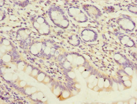
Immunohistochemical staining of human colon shows strong cytoplasmic positivity in glandular cells.
![Lane 1: Marker [kDa] 250, 130, 95, 72, 55, 36, 28, 17, 10. Lane 2: Human cell line RT-4. Lane 3: Human cell line U-251MG sp. Lane 4: Human plasma (IgG/HSA depleted). Lane 5: Human liver tissue. Lane 6: Human tonsil tissue](https://atlasantibodies.s3.amazonaws.com/images/wb/hpa044837-wb-1.jpg "Lane 1: Marker [kDa] 250, 130, 95, 72, 55, 36, 28, 17, 10. Lane 2: Human cell line RT-4. Lane 3: Human cell line U-251MG sp. Lane 4: Human plasma (IgG/HSA depleted). Lane 5: Human liver tissue. Lane 6: Human tonsil tissue")
Immunohistochemical staining of human colon shows strong cytoplasmic positivity in glandular cells.
Anti-PEX19 Antibody
HPA044837
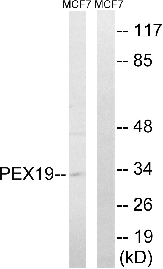
ApplicationsWestern Blot, ImmunoHistoChemistry
Product group Antibodies
ReactivityHuman, Mouse, Rat
TargetPEX19
Overview
- SupplierAtlas Antibodies
- Product NameAnti-PEX19 Antibody
- Delivery Days Customer4
- ApplicationsWestern Blot, ImmunoHistoChemistry
- CertificationResearch Use Only
- ClonalityPolyclonal
- ConjugateUnconjugated
- Gene ID5824
- Target namePEX19
- Target descriptionperoxisomal biogenesis factor 19
- Target synonymsD1S2223E, HK33, PBD12A, PMP1, PMPI, PXF, PXMP1, peroxisomal biogenesis factor 19, 33 kDa housekeeping protein, housekeeping gene, 33kD, peroxin-19, peroxisomal farnesylated protein
- HostRabbit
- IsotypeIgG
- Protein IDP40855
- Protein NamePeroxisomal biogenesis factor 19
- Scientific DescriptionRecombinant Protein Epitope Signature Tag (PrEST) antigen sequence
- ReactivityHuman, Mouse, Rat
- Storage Instruction-20°C,2°C to 8°C
- UNSPSC41116161
Datasheet
MSDS
Related products
Product group Antibodies
PEX19 AntibodyCSB-PA017802DSR2HU
ApplicationsELISA, ImmunoHistoChemistry
ReactivityHuman
TargetPEX19
- SizePrice
Product group Antibodies
Anti-PEX19 AntibodyA100808
ApplicationsWestern Blot, ELISA
ReactivityHuman
- SizePrice

Product group Antibodies
Anti-PEX19 Antibody Picoband(r)A03186-2-CARRIER-FREE
ApplicationsFlow Cytometry, Western Blot, ELISA
ReactivityHuman
TargetPEX19
- SizePrice

Product group Antibodies
Anti-PEX19 AntibodyHPA051966
ApplicationsWestern Blot, ImmunoCytoChemistry
ReactivityHuman
TargetPEX19
- SizePrice
Product group Antibodies

PEX19 Antibody (C-Terminus)LS-C368919
ApplicationsWestern Blot, ImmunoHistoChemistry, ImmunoHistoChemistry Paraffin
ReactivityHuman
TargetPEX19
- SizePrice
Product group Antibodies
PEX19 antibodyGTX110721
ApplicationsImmunoFluorescence, Western Blot, ImmunoCytoChemistry, ImmunoHistoChemistry, ImmunoHistoChemistry Paraffin
ReactivityHuman, Mouse, Rat
TargetPEX19
- SizePrice
Product group Antibodies
PEX19 Recombinant Antibody, AbBy Fluor-647 ConjugatedBSM-62360R-BF647
ApplicationsWestern Blot
ReactivityHuman, Rat
TargetPEX19
- SizePrice