


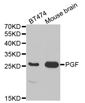
Figure 1. Western blot analysis of PGF using anti-PGF antibody (A01164-2). Electrophoresis was performed on a 5-20% SDS-PAGE gel at 70V (Stacking gel) / 90V (Resolving gel) for 2-3 hours. The sample well of each lane was loaded with 50ug of sample under reducing conditions. Lane 1: human T-47D whole cell lysate. After Electrophoresis, proteins were transferred to a Nitrocellulose membrane at 150mA for 50-90 minutes. Blocked the membrane with 5% Non-fat Milk/ TBS for 1.5 hour at RT. The membrane was incubated with rabbit anti-PGF antigen affinity purified polyclonal antibody (Catalog # A01164-2) at 0.5 microg/mL overnight at 4°C, then washed with TBS-0.1%Tween 3 times with 5 minutes each and probed with a goat anti-rabbit IgG-HRP secondary antibody at a dilution of 1:10000 for 1.5 hour at RT. The signal is developed using an Enhanced Chemiluminescent detection (ECL) kit (Catalog # EK1002) with Tanon 5200 system. A specific band was detected for PGF at approximately 25KD. The expected band size for PGF is at 25KD.
Figure 1. Western blot analysis of PGF using anti-PGF antibody (A01164-2). Electrophoresis was performed on a 5-20% SDS-PAGE gel at 70V (Stacking gel) / 90V (Resolving gel) for 2-3 hours. The sample well of each lane was loaded with 50ug of sample under reducing conditions. Lane 1: human T-47D whole cell lysate. After Electrophoresis, proteins were transferred to a Nitrocellulose membrane at 150mA for 50-90 minutes. Blocked the membrane with 5% Non-fat Milk/ TBS for 1.5 hour at RT. The membrane was incubated with rabbit anti-PGF antigen affinity purified polyclonal antibody (Catalog # A01164-2) at 0.5 microg/mL overnight at 4°C, then washed with TBS-0.1%Tween 3 times with 5 minutes each and probed with a goat anti-rabbit IgG-HRP secondary antibody at a dilution of 1:10000 for 1.5 hour at RT. The signal is developed using an Enhanced Chemiluminescent detection (ECL) kit (Catalog # EK1002) with Tanon 5200 system. A specific band was detected for PGF at approximately 25KD. The expected band size for PGF is at 25KD.
Anti-PGF Antibody Picoband(r)
A01164-2-CARRIER-FREE
ApplicationsWestern Blot, ELISA
Product group Antibodies
ReactivityHuman, Mouse
TargetPGF
Overview
- SupplierBoster Bio
- Product NameAnti-PGF Antibody Picoband(r)
- Delivery Days Customer9
- ApplicationsWestern Blot, ELISA
- CertificationResearch Use Only
- ClonalityPolyclonal
- Concentration500 ug/ml
- Gene ID5228
- Target namePGF
- Target descriptionplacental growth factor
- Target synonymsD12S1900, PGFL, PIGF, PLGF, PlGF-2, SHGC-10760, placenta growth factor, placental growth factor, vascular endothelial growth factor-related protein
- HostRabbit
- IsotypeIgG
- Protein IDP49763
- Protein NamePlacenta growth factor
- Scientific DescriptionBoster Bio Anti-PGF Antibody Picoband® catalog # A01164-2. Tested in ELISA, WB applications. This antibody reacts with Human, Mouse. The brand Picoband indicates this is a premium antibody that guarantees superior quality, high affinity, and strong signals with minimal background in Western blot applications. Only our best-performing antibodies are designated as Picoband, ensuring unmatched performance.
- ReactivityHuman, Mouse
- Storage Instruction-20°C,2°C to 8°C
- UNSPSC12352203
Related products
Product group Antibodies
Anti-PGF AntibodyA30493
ApplicationsWestern Blot, ImmunoHistoChemistry
ReactivityHuman, Mouse, Rat
- SizePrice
Product group Antibodies
Anti-PGF Antibody130-10284
ApplicationsWestern Blot, ELISA
ReactivityHuman
TargetPGF
- SizePrice
Product group Antibodies
PLGF Polyclonal AntibodyBS-0281R
ApplicationsImmunoFluorescence, Western Blot, ELISA, ImmunoCytoChemistry, ImmunoHistoChemistry, ImmunoHistoChemistry Frozen, ImmunoHistoChemistry Paraffin
ReactivityHuman
TargetPGF
- SizePrice
Product group Antibodies
PGF AntibodyCSB-PA06959A0RB
ApplicationsELISA
ReactivityHuman
TargetPGF
- SizePrice
Product group Antibodies
ApplicationsImmunoPrecipitation, Western Blot, ImmunoCytoChemistry, ImmunoHistoChemistry
TargetPGF
- SizePrice
Product group Antibodies

ApplicationsWestern Blot, ELISA
ReactivityHuman
TargetPGF
- SizePrice
![Sandwich ELISA detection of recombinant Human PLGF protein, His tag (GTX138450-pro) using antibodies as below. Capture: PLGF antibody [GT57] (GTX641446) (5 μg/mL) Detection: PLGF antibody [HL2682] (GTX639346) (1 μg/mL)](https://www.genetex.com/upload/website/prouct_img/normal/GTX639346/GTX639346_T-45222_20241220_ELISA_PAIR_24123021_606.webp)
Product group Antibodies
PLGF antibody [HL2682]GTX639346
ApplicationsELISA
ReactivityHuman
TargetPGF
- SizePrice
Product group Antibodies
Anti-PDE12 AntibodyCAB17270
ApplicationsImmunoFluorescence, Western Blot, ELISA, ImmunoCytoChemistry
ReactivityMouse
TargetPGF
- SizePrice