


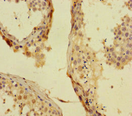
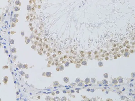
Immunohistochemical staining of human pancreas shows strong nuclear positivity in islet cells.

Immunohistochemical staining of human pancreas shows strong nuclear positivity in islet cells.
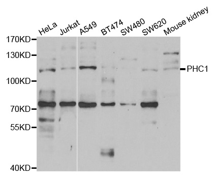
Anti-PHC1 Antibody
HPA006973
ApplicationsImmunoCytoChemistry, ImmunoHistoChemistry
Product group Antibodies
ReactivityHuman
TargetPHC1
Overview
- SupplierAtlas Antibodies
- Product NameAnti-PHC1 Antibody
- Delivery Days Customer4
- ApplicationsImmunoCytoChemistry, ImmunoHistoChemistry
- CertificationResearch Use Only
- ClonalityPolyclonal
- ConjugateUnconjugated
- Gene ID1911
- Target namePHC1
- Target descriptionpolyhomeotic homolog 1
- Target synonymsEDR1, HPH1, MCPH11, RAE28, polyhomeotic-like protein 1, early development regulator 1 (homolog of polyhomeotic 1), early development regulatory protein 1, polyhomeotic-like 1
- HostRabbit
- IsotypeIgG
- Protein IDP78364
- Protein NamePolyhomeotic-like protein 1
- Scientific DescriptionRecombinant Protein Epitope Signature Tag (PrEST) antigen sequence
- ReactivityHuman
- Storage Instruction-20°C,2°C to 8°C
- UNSPSC41116161
Datasheet
MSDS
Related products
Product group Antibodies
PHC1 AntibodyCSB-PA017891HA01HU
ApplicationsELISA, ImmunoHistoChemistry
ReactivityHuman
TargetPHC1
- SizePrice

Product group Antibodies
Anti-PHC1 Antibody Picoband(r)A06437-3-CARRIER-FREE
ApplicationsImmunoFluorescence, Western Blot, ELISA, ImmunoCytoChemistry
ReactivityHuman
TargetPHC1
- SizePrice
Product group Antibodies
Anti-PHC1 AntibodyA31087
ApplicationsWestern Blot
ReactivityHuman, Mouse, Rat
- SizePrice
Product group Antibodies

PHC1 / EDR1 AntibodyLS-C335607
ApplicationsWestern Blot, ImmunoHistoChemistry
ReactivityHuman, Mouse, Rat
TargetPHC1
- SizePrice
Product group Antibodies
PHC1 antibodyGTX32784
ApplicationsWestern Blot, ImmunoHistoChemistry, ImmunoHistoChemistry Paraffin
ReactivityHuman, Mouse, Rat
TargetPHC1
- SizePrice
Product group Antibodies
Anti-PHC1 Antibody144-05843
ApplicationsWestern Blot, ImmunoHistoChemistry
ReactivityHuman, Mouse, Rat
TargetPHC1
- SizePrice
Product group Antibodies
PHC1 Polyclonal AntibodyBS-12302R
ApplicationsImmunoFluorescence, ELISA, ImmunoCytoChemistry, ImmunoHistoChemistry, ImmunoHistoChemistry Frozen, ImmunoHistoChemistry Paraffin
ReactivityBovine, Canine, Human, Mouse, Porcine, Rabbit, Rat, Sheep
TargetPHC1
- SizePrice