

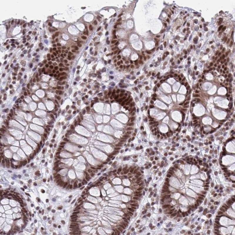
Immunohistochemical staining of human colon shows strong nuclear positivity in glandular cells.

Immunohistochemical staining of human colon shows strong nuclear positivity in glandular cells.
Anti-PHF10 Antibody
HPA055649
ApplicationsImmunoCytoChemistry, ImmunoHistoChemistry
Product group Antibodies
ReactivityHuman
TargetPHF10
Overview
- SupplierAtlas Antibodies
- Product NameAnti-PHF10 Antibody
- Delivery Days Customer4
- ApplicationsImmunoCytoChemistry, ImmunoHistoChemistry
- CertificationResearch Use Only
- ClonalityPolyclonal
- ConjugateUnconjugated
- Gene ID55274
- Target namePHF10
- Target descriptionPHD finger protein 10
- Target synonymsBAF45A, SMARCG4, XAP135, PHD finger protein 10, BRG1-associated factor 45a, BRG1-associated factor, 45-KD, A, PHD zinc finger protein XAP135
- HostRabbit
- IsotypeIgG
- Protein IDQ8WUB8
- Protein NamePHD finger protein 10
- Scientific DescriptionRecombinant Protein Epitope Signature Tag (PrEST) antigen sequence
- ReactivityHuman
- Storage Instruction-20°C,2°C to 8°C
- UNSPSC41116161
Datasheet
MSDS
Related products

Product group Antibodies
Anti-PHF10 Antibody Picoband(r)A08195-1-CARRIER-FREE
ApplicationsFlow Cytometry, Western Blot, ELISA
ReactivityHuman, Mouse, Rat
TargetPHF10
- SizePrice

Product group Antibodies
Anti-PHF10 AntibodyA89684
ApplicationsWestern Blot
ReactivityHuman, Mouse
- SizePrice
Product group Antibodies

PHF10 AntibodyLS-C750437
ApplicationsWestern Blot
ReactivityHuman, Mouse
TargetPHF10
- SizePrice
Product group Antibodies
PHF10 Polyclonal AntibodyCAC12915
ApplicationsImmunoFluorescence, Western Blot, ELISA, ImmunoHistoChemistry
TargetPHF10
- SizePrice
Product group Antibodies
PHF10 AntibodyCSB-PA017898LA01HU
ApplicationsImmunoFluorescence, Western Blot, ELISA, ImmunoHistoChemistry
ReactivityHuman
TargetPHF10
- SizePrice
Product group Antibodies
PHF10 antibodyGTX116314
ApplicationsImmunoFluorescence, Western Blot, ImmunoCytoChemistry, ImmunoHistoChemistry, ImmunoHistoChemistry Paraffin
ReactivityHuman, Mouse, Rat
TargetPHF10
- SizePrice