


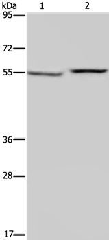
Figure 1. Western blot analysis of PHGDH using anti-PHGDH antibody (A03355-1). Electrophoresis was performed on a 5-20% SDS-PAGE gel at 70V (Stacking gel) / 90V (Resolving gel) for 2-3 hours. The sample well of each lane was loaded with 30 ug of sample under reducing conditions. Lane 1: human U251 whole cell lysates, Lane 2: human Jurkat whole cell lysates, Lane 3: human HEL whole cell lysates, Lane 4: human K562 whole cell lysates, Lane 5: rat liver tissue lysates, Lane 6: rat kidney tissue lysates, Lane 7: mouse liver tissue lysates, Lane 8: mouse kidney tissue lysates. After electrophoresis, proteins were transferred to a nitrocellulose membrane at 150 mA for 50-90 minutes. Blocked the membrane with 5% non-fat milk/TBS for 1.5 hour at RT. The membrane was incubated with rabbit anti-PHGDH antigen affinity purified polyclonal antibody (A03355-1) at 0.5 microg/mL overnight at 4°C, then washed with TBS-0.1%Tween 3 times with 5 minutes each and probed with a goat anti-rabbit IgG-HRP secondary antibody at a dilution of 1:5000 for 1.5 hour at RT. The signal is developed using an Enhanced Chemiluminescent detection (ECL) kit (Catalog # EK1002) with Tanon 5200 system. A specific band was detected for PHGDH at approximately 57 kDa. The expected band size for PHGDH is at 57 kDa.
. PHGDH was detected in a paraffin-embedded section of human breast cancer tissue. Heat mediated antigen retrieval was performed in EDTA buffer (pH 8.0, epitope retrieval solution). The tissue section was blocked with 10% goat serum. The tissue section was then incubated with 2 microg/ml rabbit anti-PHGDH Antibody (A03355-1) overnight at 4°C. Peroxidase Conjugated Goat Anti-rabbit IgG was used as secondary antibody and incubated for 30 minutes at 37°C. The tissue section was developed using HRP Conjugated Rabbit IgG Super Vision Assay Kit (Catalog # SV0002) with DAB as the chromogen.")
. PHGDH was detected in an immunocytochemical section of U2OS cells. Enzyme antigen retrieval was performed using IHC enzyme antigen retrieval reagent (AR0022) for 15 mins. The cells were blocked with 10% goat serum. And then incubated with 4 microg/mL rabbit anti-PHGDH Antibody (A03355-1) overnight at 4°C. DyLight®488 Conjugated Goat Anti-Rabbit IgG (BA1127) was used as secondary antibody at 1:500 dilution and incubated for 30 minutes at 37°C. The section was counterstained with DAPI. Visualize using a fluorescence microscope and filter sets appropriate for the label used.")
. Overlay histogram showing PC-3 cells stained with A03355-1 (Blue line). To facilitate intracellular staining, cells were fixed with 4% paraformaldehyde and permeabilized with permeabilization buffer. The cells were blocked with 10% normal goat serum. And then incubated with rabbit anti-PHGDH Antibody (A03355-1, 1 microg/1x106 cells) for 30 min at 20°C. DyLight®488 conjugated goat anti-rabbit IgG (BA1127, 5-10 microg/1x106 cells) was used as secondary antibody for 30 minutes at 20°C. Isotype control antibody (Green line) was rabbit IgG (1 microg/1x106) used under the same conditions. Unlabelled sample without incubation with primary antibody and secondary antibody (Red line) was used as a blank control.")
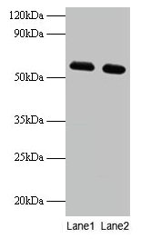
Figure 1. Western blot analysis of PHGDH using anti-PHGDH antibody (A03355-1). Electrophoresis was performed on a 5-20% SDS-PAGE gel at 70V (Stacking gel) / 90V (Resolving gel) for 2-3 hours. The sample well of each lane was loaded with 30 ug of sample under reducing conditions. Lane 1: human U251 whole cell lysates, Lane 2: human Jurkat whole cell lysates, Lane 3: human HEL whole cell lysates, Lane 4: human K562 whole cell lysates, Lane 5: rat liver tissue lysates, Lane 6: rat kidney tissue lysates, Lane 7: mouse liver tissue lysates, Lane 8: mouse kidney tissue lysates. After electrophoresis, proteins were transferred to a nitrocellulose membrane at 150 mA for 50-90 minutes. Blocked the membrane with 5% non-fat milk/TBS for 1.5 hour at RT. The membrane was incubated with rabbit anti-PHGDH antigen affinity purified polyclonal antibody (A03355-1) at 0.5 microg/mL overnight at 4°C, then washed with TBS-0.1%Tween 3 times with 5 minutes each and probed with a goat anti-rabbit IgG-HRP secondary antibody at a dilution of 1:5000 for 1.5 hour at RT. The signal is developed using an Enhanced Chemiluminescent detection (ECL) kit (Catalog # EK1002) with Tanon 5200 system. A specific band was detected for PHGDH at approximately 57 kDa. The expected band size for PHGDH is at 57 kDa.
Anti-PHGDH Antibody Picoband(r)
A03355-1-CARRIER-FREE
ApplicationsFlow Cytometry, ImmunoFluorescence, ImmunoPrecipitation, Western Blot, ELISA, ImmunoCytoChemistry, ImmunoHistoChemistry
Product group Antibodies
ReactivityHuman, Mouse, Rat
TargetPHGDH
Overview
- SupplierBoster Bio
- Product NameAnti-PHGDH Antibody Picoband(r)
- Delivery Days Customer9
- ApplicationsFlow Cytometry, ImmunoFluorescence, ImmunoPrecipitation, Western Blot, ELISA, ImmunoCytoChemistry, ImmunoHistoChemistry
- CertificationResearch Use Only
- ClonalityPolyclonal
- Concentration500 ug/ml
- Gene ID26227
- Target namePHGDH
- Target descriptionphosphoglycerate dehydrogenase
- Target synonyms3-PGDH, 3PGDH, HEL-S-113, NLS, NLS1, PDG, PGAD, PGD, PGDH, PHGDHD, SERA, D-3-phosphoglycerate dehydrogenase, 2-oxoglutarate reductase, 3-phosphoglycerate dehydrogenase, epididymis secretory protein Li 113, malate dehydrogenase
- HostRabbit
- IsotypeIgG
- Protein IDO43175
- Protein NameD-3-phosphoglycerate dehydrogenase
- Scientific DescriptionBoster Bio Anti-PHGDH Antibody Picoband® catalog # A03355-1. Tested in ELISA, Flow Cytometry, IP, IF, IHC, ICC, WB applications. This antibody reacts with Human, Mouse, Rat. The brand Picoband indicates this is a premium antibody that guarantees superior quality, high affinity, and strong signals with minimal background in Western blot applications. Only our best-performing antibodies are designated as Picoband, ensuring unmatched performance.
- ReactivityHuman, Mouse, Rat
- Storage Instruction-20°C,2°C to 8°C
- UNSPSC12352203
Related products
Product group Antibodies
Anti-PHGDH Antibody144-10461
ApplicationsWestern Blot, ImmunoHistoChemistry
ReactivityHuman, Mouse, Rat
TargetPHGDH
- SizePrice

Product group Antibodies
Anti-PHGDH AntibodyAMAB90786
ApplicationsWestern Blot, ImmunoCytoChemistry, ImmunoHistoChemistry
ReactivityHuman
TargetPHGDH
- SizePrice
Product group Antibodies
Anti-PHGDH AntibodyA43254
ApplicationsWestern Blot
ReactivityHuman, Mouse
- SizePrice
Product group Antibodies
PHGDH Polyclonal AntibodyCAC14035
ApplicationsImmunoFluorescence, Western Blot, ELISA, ImmunoHistoChemistry
TargetPHGDH
- SizePrice
Product group Antibodies
PHGDH AntibodyCSB-PA03255A0RB
ApplicationsImmunoFluorescence, Western Blot, ELISA, ImmunoHistoChemistry
ReactivityHuman
TargetPHGDH
- SizePrice
Product group Antibodies
PHGDH Polyclonal AntibodyBS-2970R
ApplicationsImmunoFluorescence, Western Blot, ELISA, ImmunoCytoChemistry, ImmunoHistoChemistry, ImmunoHistoChemistry Frozen, ImmunoHistoChemistry Paraffin
ReactivityBovine, Canine, Equine, Human, Mouse, Porcine, Rabbit, Rat, Sheep
TargetPHGDH
- SizePrice
![Immunoprecipitation of PHGDH protein from 293T whole cell extracts using 5 μg of PHGDH antibody [N1N2], N-term (GTX101948). Western blot analysis was performed using PHGDH antibody [N1N2], N-term (GTX101948). EasyBlot anti-Rabbit IgG (GTX221666-01) was used as a secondary reagent.](https://www.genetex.com/upload/website/prouct_img/normal/GTX101948/GTX101948_39834_20150407_IP_w_23060100_291.webp)
Product group Antibodies
PHGDH antibody [N1N2], N-termGTX101948
ApplicationsImmunoFluorescence, ImmunoPrecipitation, Western Blot, ImmunoCytoChemistry, ImmunoHistoChemistry, ImmunoHistoChemistry Frozen, ImmunoHistoChemistry Paraffin
ReactivityHuman, Mouse, Rat
TargetPHGDH
- SizePrice
Product group Antibodies

PHGDH AntibodyLS-C497170
ApplicationsWestern Blot, ImmunoHistoChemistry
ReactivityHuman, Mouse, Rat
TargetPHGDH
- SizePrice