

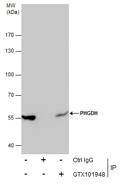
Immunoprecipitation of PHGDH protein from 293T whole cell extracts using 5 μg of PHGDH antibody [N1N2], N-term (GTX101948). Western blot analysis was performed using PHGDH antibody [N1N2], N-term (GTX101948). EasyBlot anti-Rabbit IgG (GTX221666-01) was used as a secondary reagent.
 antibody at 1:500 dilution.
Antigen Retrieval: Trilogy? (EDTA based, pH 8.0) buffer, 15min")
![PHGDH antibody [N1N2], N-term detects PHGDH protein by immunohistochemical analysis. Sample: Frozen-sectioned rat E13.5 brain. Green: PHGDH stained by PHGDH antibody [N1N2], N-term (GTX101948) diluted at 1:250. Blue: Fluoroshield with DAPI (GTX30920).](https://www.genetex.com/upload/website/prouct_img/normal/GTX101948/GTX101948_39834_20171106_IHC-Fr_R_w_23060100_932.webp "PHGDH antibody [N1N2], N-term detects PHGDH protein by immunohistochemical analysis. Sample: Frozen-sectioned rat E13.5 brain. Green: PHGDH stained by PHGDH antibody [N1N2], N-term (GTX101948) diluted at 1:250. Blue: Fluoroshield with DAPI (GTX30920).")
 A: Mouse brain 7.5% SDS PAGE GTX101948 diluted at 1:1000")
 A:293T B:A431(GTX27909) C:HeLa S3(GTX14654) 7.5% SDS PAGE GTX101948 diluted at 1:1000")
 antibody at 1:500 dilution.")
![Wild-type (WT) and PHGDH knockout (KO) HeLa cell extracts (30 μg) were separated by 7.5% SDS-PAGE, and the membrane was blotted with PHGDH antibody [N1N2], N-term (GTX101948) diluted at 1:500. The HRP-conjugated anti-rabbit IgG antibody (GTX213110-01) was used to detect the primary antibody.](https://www.genetex.com/upload/website/prouct_img/normal/GTX101948/GTX101948_39834_20170615_WB_KO_watermark_w_23060100_769.webp "Wild-type (WT) and PHGDH knockout (KO) HeLa cell extracts (30 μg) were separated by 7.5% SDS-PAGE, and the membrane was blotted with PHGDH antibody [N1N2], N-term (GTX101948) diluted at 1:500. The HRP-conjugated anti-rabbit IgG antibody (GTX213110-01) was used to detect the primary antibody.")
![PHGDH antibody [N1N2], N-term detects PHGDH protein at cytoplasm on mouse pancreas by immunohistochemical analysis. Sample: Paraffin-embedded mouse pancreas. PHGDH antibody [N1N2], N-term (GTX101948) diluted at 1:500.
Antigen Retrieval: Trilogy? (EDTA based, pH 8.0) buffer, 15min](https://www.genetex.com/upload/website/prouct_img/normal/GTX101948/GTX101948_39834_20150116_IHC_M_w_23060100_737.webp "PHGDH antibody [N1N2], N-term detects PHGDH protein at cytoplasm on mouse pancreas by immunohistochemical analysis. Sample: Paraffin-embedded mouse pancreas. PHGDH antibody [N1N2], N-term (GTX101948) diluted at 1:500.
Antigen Retrieval: Trilogy? (EDTA based, pH 8.0) buffer, 15min")
Immunoprecipitation of PHGDH protein from 293T whole cell extracts using 5 μg of PHGDH antibody [N1N2], N-term (GTX101948). Western blot analysis was performed using PHGDH antibody [N1N2], N-term (GTX101948). EasyBlot anti-Rabbit IgG (GTX221666-01) was used as a secondary reagent.
PHGDH antibody [N1N2], N-term
GTX101948
ApplicationsImmunoFluorescence, ImmunoPrecipitation, Western Blot, ImmunoCytoChemistry, ImmunoHistoChemistry, ImmunoHistoChemistry Frozen, ImmunoHistoChemistry Paraffin
Product group Antibodies
ReactivityHuman, Mouse, Rat
TargetPHGDH
Overview
- SupplierGeneTex
- Product NamePHGDH antibody [N1N2], N-term
- Delivery Days Customer9
- Application Supplier NoteWB: 1:500-1:3000. ICC/IF: 1:100-1:1000. IHC-P: 1:100-1:1000. IHC-Fr: 1:100-1:1000. IP: 1:100-1:500. *Optimal dilutions/concentrations should be determined by the researcher.Not tested in other applications.
- ApplicationsImmunoFluorescence, ImmunoPrecipitation, Western Blot, ImmunoCytoChemistry, ImmunoHistoChemistry, ImmunoHistoChemistry Frozen, ImmunoHistoChemistry Paraffin
- CertificationResearch Use Only
- ClonalityPolyclonal
- Concentration0.87 mg/ml
- ConjugateUnconjugated
- Gene ID26227
- Target namePHGDH
- Target descriptionphosphoglycerate dehydrogenase
- Target synonyms3-PGDH, 3PGDH, HEL-S-113, NLS, NLS1, PDG, PGAD, PGD, PGDH, PHGDHD, SERA, D-3-phosphoglycerate dehydrogenase, 2-oxoglutarate reductase, 3-phosphoglycerate dehydrogenase, epididymis secretory protein Li 113, malate dehydrogenase
- HostRabbit
- IsotypeIgG
- Protein IDO43175
- Protein NameD-3-phosphoglycerate dehydrogenase
- Scientific Description3-Phosphoglycerate dehydrogenase (PHGDH; EC 1.1.1.95) catalyzes the transition of 3-phosphoglycerate into 3-phosphohydroxypyruvate, which is the first and rate-limiting step in the phosphorylated pathway of serine biosynthesis, using NAD+/NADH as a cofactor.[supplied by OMIM]
- ReactivityHuman, Mouse, Rat
- Storage Instruction-20°C or -80°C,2°C to 8°C
- UNSPSC41116161
Datasheet
Related products

Product group Antibodies
Anti-PHGDH Antibody Picoband(r)A03355-1-CARRIER-FREE
ApplicationsFlow Cytometry, ImmunoFluorescence, ImmunoPrecipitation, Western Blot, ELISA, ImmunoCytoChemistry, ImmunoHistoChemistry
ReactivityHuman, Mouse, Rat
TargetPHGDH
- SizePrice
Product group Antibodies
Anti-PHGDH Antibody144-10461
ApplicationsWestern Blot, ImmunoHistoChemistry
ReactivityHuman, Mouse, Rat
TargetPHGDH
- SizePrice

Product group Antibodies
Anti-PHGDH AntibodyAMAB90786
ApplicationsWestern Blot, ImmunoCytoChemistry, ImmunoHistoChemistry
ReactivityHuman
TargetPHGDH
- SizePrice
Product group Antibodies
Anti-PHGDH AntibodyA43254
ApplicationsWestern Blot
ReactivityHuman, Mouse
- SizePrice
Product group Antibodies
PHGDH Polyclonal AntibodyCAC14035
ApplicationsImmunoFluorescence, Western Blot, ELISA, ImmunoHistoChemistry
TargetPHGDH
- SizePrice
Product group Antibodies
PHGDH AntibodyCSB-PA03255A0RB
ApplicationsImmunoFluorescence, Western Blot, ELISA, ImmunoHistoChemistry
ReactivityHuman
TargetPHGDH
- SizePrice
Product group Antibodies
PHGDH Polyclonal AntibodyBS-2970R
ApplicationsImmunoFluorescence, Western Blot, ELISA, ImmunoCytoChemistry, ImmunoHistoChemistry, ImmunoHistoChemistry Frozen, ImmunoHistoChemistry Paraffin
ReactivityBovine, Canine, Equine, Human, Mouse, Porcine, Rabbit, Rat, Sheep
TargetPHGDH
- SizePrice
![Wild-type (WT) and PHGDH knockout (KO) HeLa cell extracts (30 μg) were separated by 7.5% SDS-PAGE, and the membrane was blotted with PHGDH antibody [N3C2], Internal (GTX101949) diluted at 1:1000. The HRP-conjugated anti-rabbit IgG antibody (GTX213110-01) was used to detect the primary antibody.](https://www.genetex.com/upload/website/prouct_img/normal/GTX101949/GTX101949_40604_20170818_WB_KO_watermark_w_23060100_485.webp)
Product group Antibodies
PHGDH antibody [N3C2], InternalGTX101949
ApplicationsWestern Blot, ImmunoHistoChemistry, ImmunoHistoChemistry Frozen, ImmunoHistoChemistry Paraffin
ReactivityHuman, Rat
TargetPHGDH
- SizePrice
Product group Antibodies

PHGDH AntibodyLS-C497170
ApplicationsWestern Blot, ImmunoHistoChemistry
ReactivityHuman, Mouse, Rat
TargetPHGDH
- SizePrice
Product group Antibodies
PHGDH antibodyGTX64499
ApplicationsImmunoFluorescence, Western Blot, ImmunoCytoChemistry
ReactivityHuman, Mouse, Rat
TargetPHGDH
- SizePrice